Vararia investiens (Schwein.) P. Karst.
Basidiomycota: Agaricomycotina: Agaricomycetes: Russulales: Lachnocladiaceae: Vararia ![]()
![]()
Introduction
The genus Vararia, which contains about 60 species, is common in the subtropics and tropics but less diverse in temperate areas (Karasiński 2010). Of the temperate species, Vararia investiens seems to be the most widespread and frequently encountered. As far as crusts go, it is actually really well documented with 967 records in MyCoPortal (accessed December 5, 2020). I personally observed this species many times before finally collecting one for study. It appears to be prevalent on the underside of small deciduous branches and sticks, recognizable by its distinct yellow color and velutinous aspect. Any doubts about ID are quickly dispelled with microscopy — the stunning dichohyphidia and prominently pointed ellipsoid-fusoid basidiospores are quite unique.
Vararia basidiomata have an interesting development whereby the basidia and cystidia originate deep within a blanketing hyphidial hymenium and elongate through it, but are constantly overgrown by expanding hyphal elements. As a result, basidia and basidiospores are found embedded within the basidiocarp at all levels and are difficult to discern among the jungle of needley dichohyphae (Lentz & McKay 1965).
Description
Ecology: Saprotroph on the underside of hardwood sticks, common and cosmopolitan.
Basidiocarp: Effused, straw yellow to golden in color, even to tuberculate, membranaceous to chalky-crustaceous (reminiscent of felt, in my opinion), growing over and agglutinating small debris to the substrate, margin abrupt.
Chemical reactions: Ammonia and KOH result in a reddish brown color; iron salts do not absorb (functionally negative).
Spore print: NA
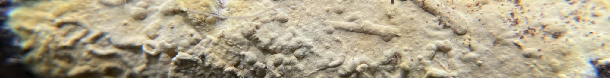
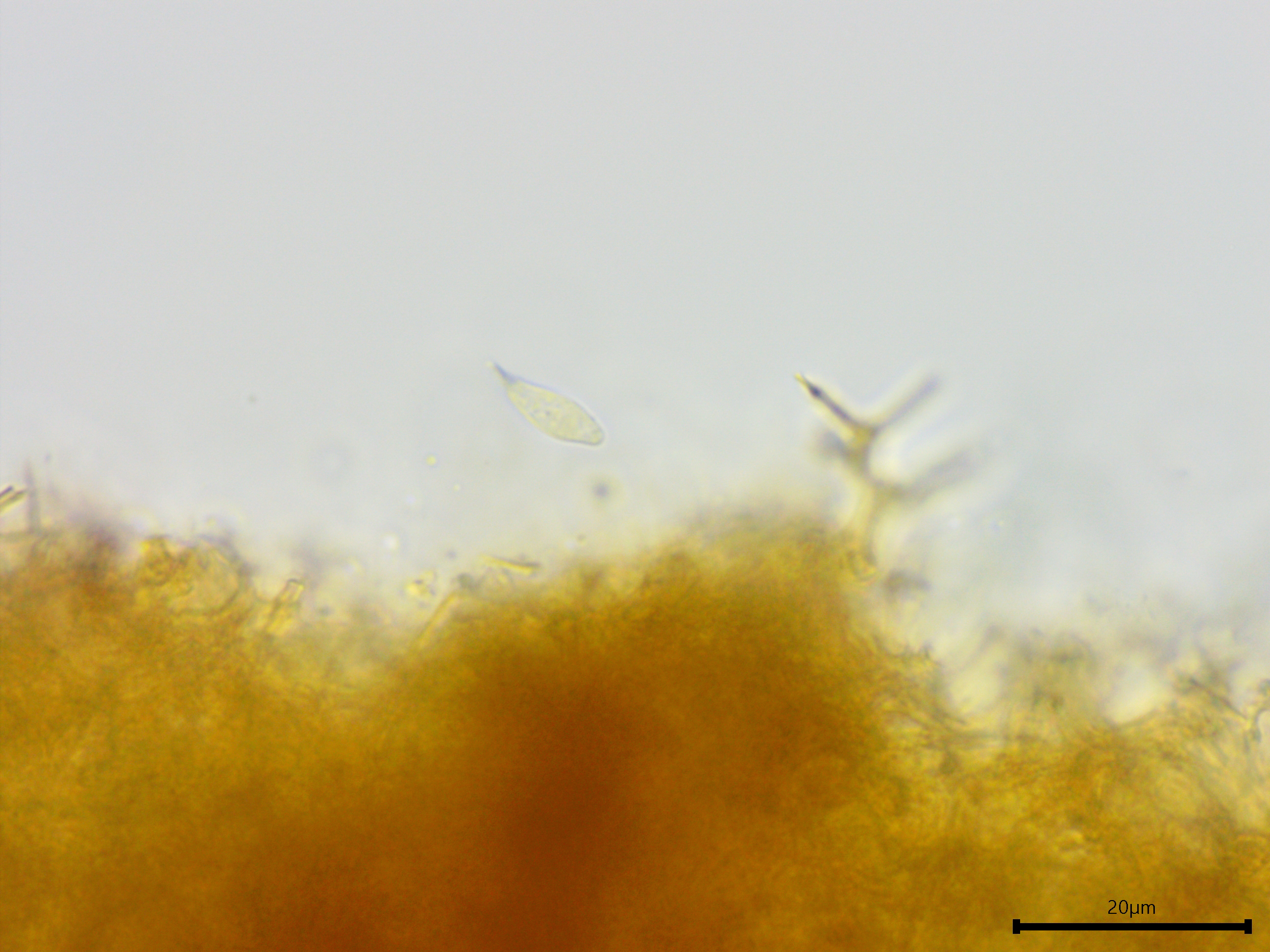
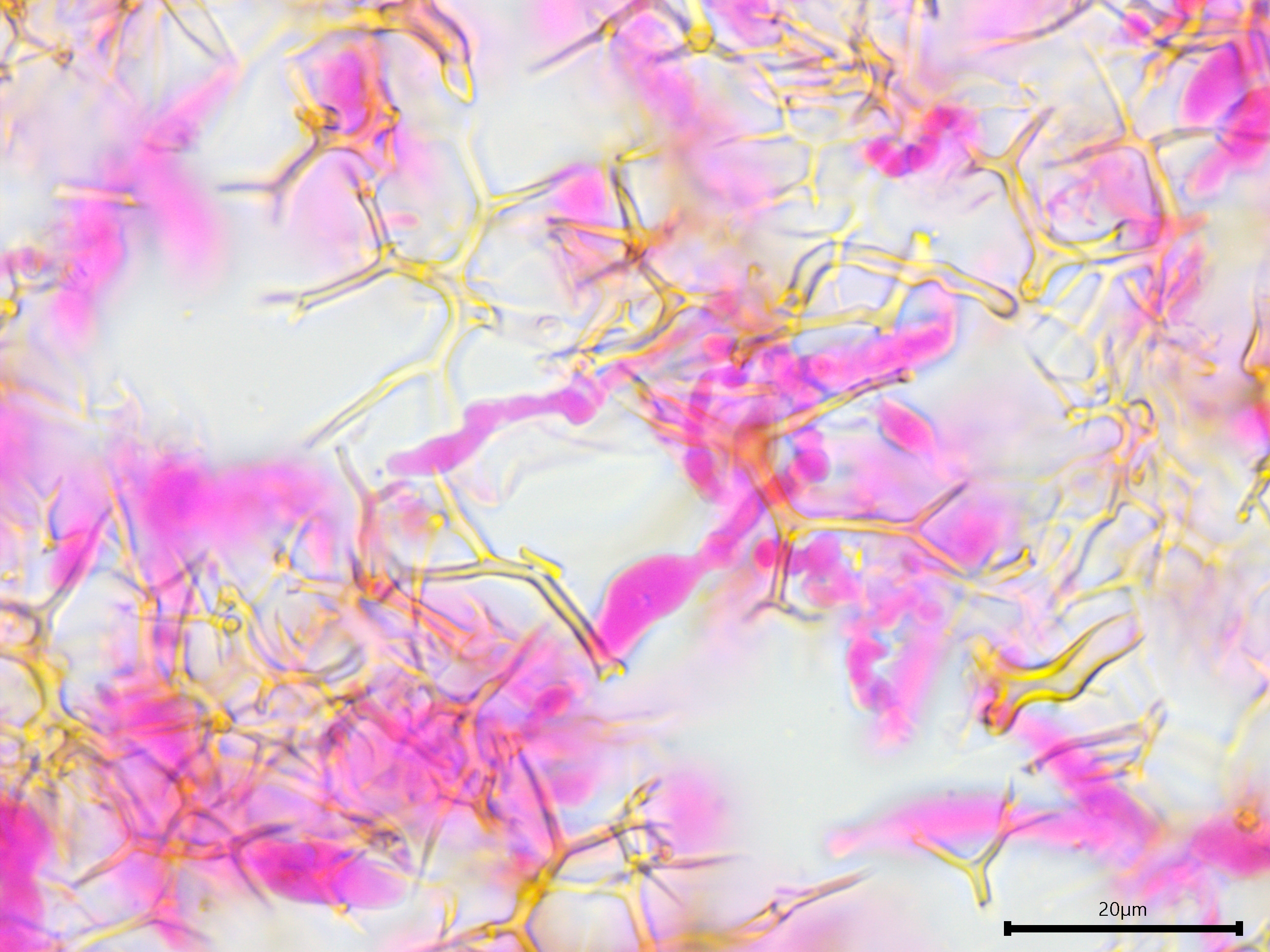
Hyphal system: Dimitic; generative hyphae with difficult-to-discern clamp connections, embedded deep within a thicket of acicular (needle-like) dichohyphae or dichohyphidia; dichohyphae thick-walled, weakly dextrinoid, yellowish in KOH, profusely branched, extending from the substrate to the hymenium and beyond, seemingly constituting the entire subiculum, width (1.9) 1.9–2.8 (3.2) µm (n = 10); I observed a few enormous and thick-walled seta-like dichohyphida with one or more central hyphae running the interior length, a characteristic mentioned briefly by Lentz and McKay (1965).
Basidia: Difficult to find within the thicket of dichohyphidia; utriform with four sterigmata.
Basidiospores: Narrowly cylindrical, fusoid or pip-shaped (like an apple seed), more accurately geniculately fusoid-ellipsoid; smooth, thin-walled, hyaline, inamyloid, with a prominently pointed (pseudo)septate amyloid tip (deemed a "suprahilar plage" or "plaque") that does not stain with phloxine B; length (8.8) 9.6–11.5 (12.5) µm, width (3.3) 3.6–4.3 (5) µm, x̄ = 10.6 ✕ 3.9 µm, Q (2.2) 2.4-3 (3.3), x̄ = 2.7 (n = 30).
Sterile structures: Gloeocystidia present, about 5–7 µm wide, up to 50 µm long.
Sequences: ITS rDNA (MZ919258, OP508177), LSU rDNA (OP596486).
Notes: All measurements taken in 5% KOH stained with phloxine B. The basidiospores are a bit wider than what is reported for this species by Bernicchia and Gorjón (2010), but more consistent with the width reported by Karasiński (2010). According to the latter study, the pip-shaped spores with their prominent "suprahilar" extension is distinct among Vararia in temperate areas (but not the tropics or subtropics), making northern specimens of this species easily identifiable with microscopy.
Specimens Analyzed
ACD0382, iNat62862821; 17 October 2020; Nan Weston Preserve, Washtenaw Co., MI, USA, 42.1856 -84.1111; leg. & det. Alden C. Dirks, ref. Bernicchia & Gorjón (2010); University of Michigan Fungarium MICH 352290.
ACD0543, iNat130261019; 10 August 2022; French Creek State Park, Chester Co., PA, USA, 40.2051 -75.7361; leg. & det. Alden C. Dirks, ref. Bernicchia & Gorjón (2010); University of Michigan Fungarium ——.
References
Bernicchia, A. & Gorjón, S.P. (2010). Fungi Europaei, Volume 12: Corticiaceae s.l. Massimo Candusso, Italia.
Karasiński, D. (2010). Polish resupinate Russulales: The genus Vararia. Acta Mycologica, 45(1), 45–56.
Lentz, P. L., & McKay, H. H. (1965). Delineations of forest fungi: Morphology and relationships of Vararia. Mycopathologia et Mycologia Applicata, 29(1–2), 1–25.
Links
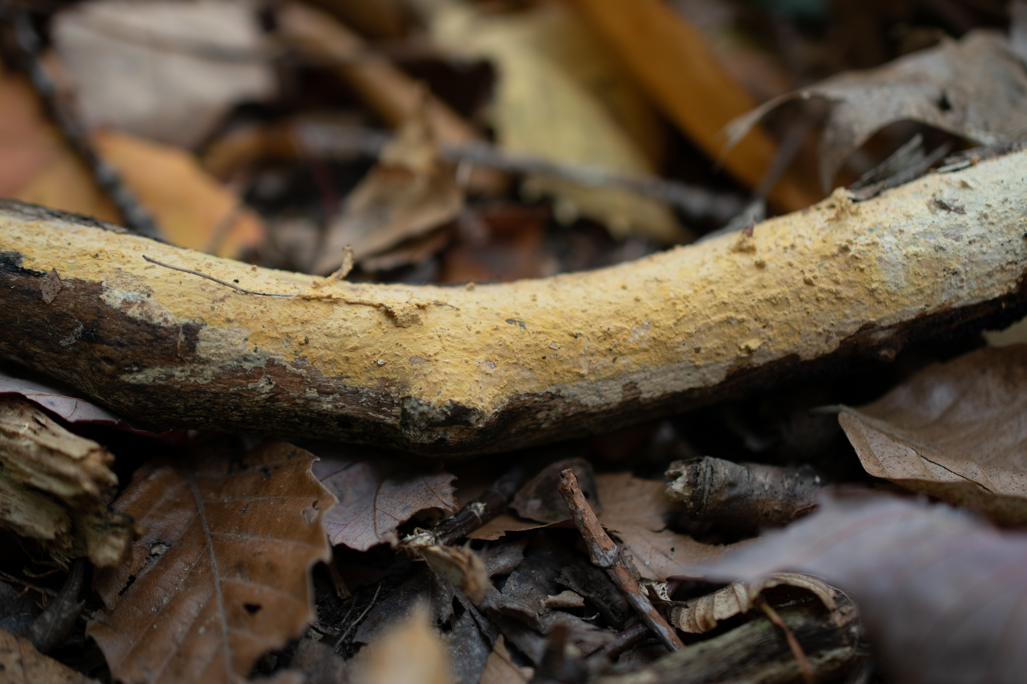
Vararia investiens is a straw yellow to golden crust.
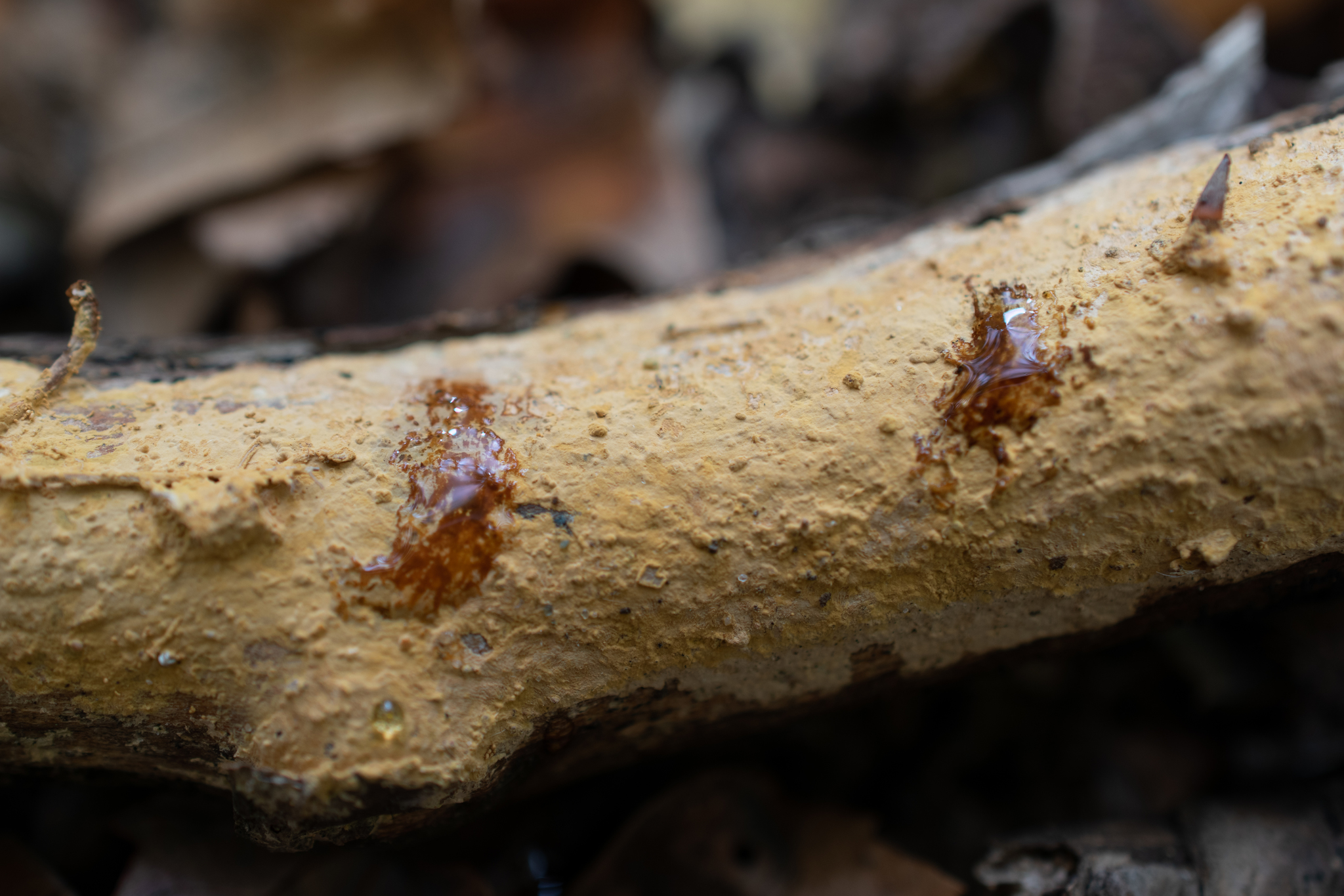
Ammonia and potassium hydroxide turn the hymenium a reddish brown color.
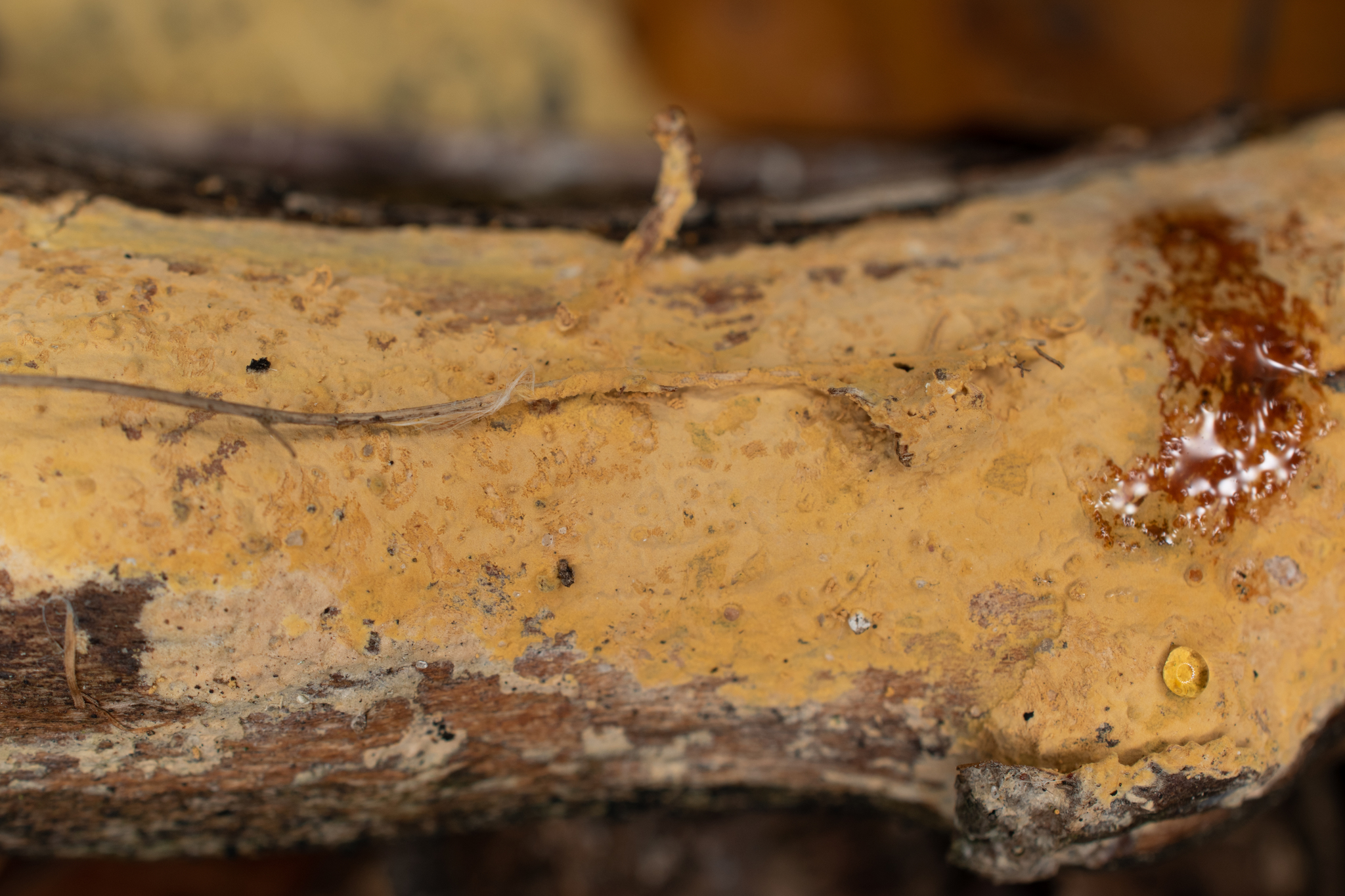
Close up, the texture is even with scattered warts. The fungus has grown over debris and small twigs, incorporating them into the basidiocarp.
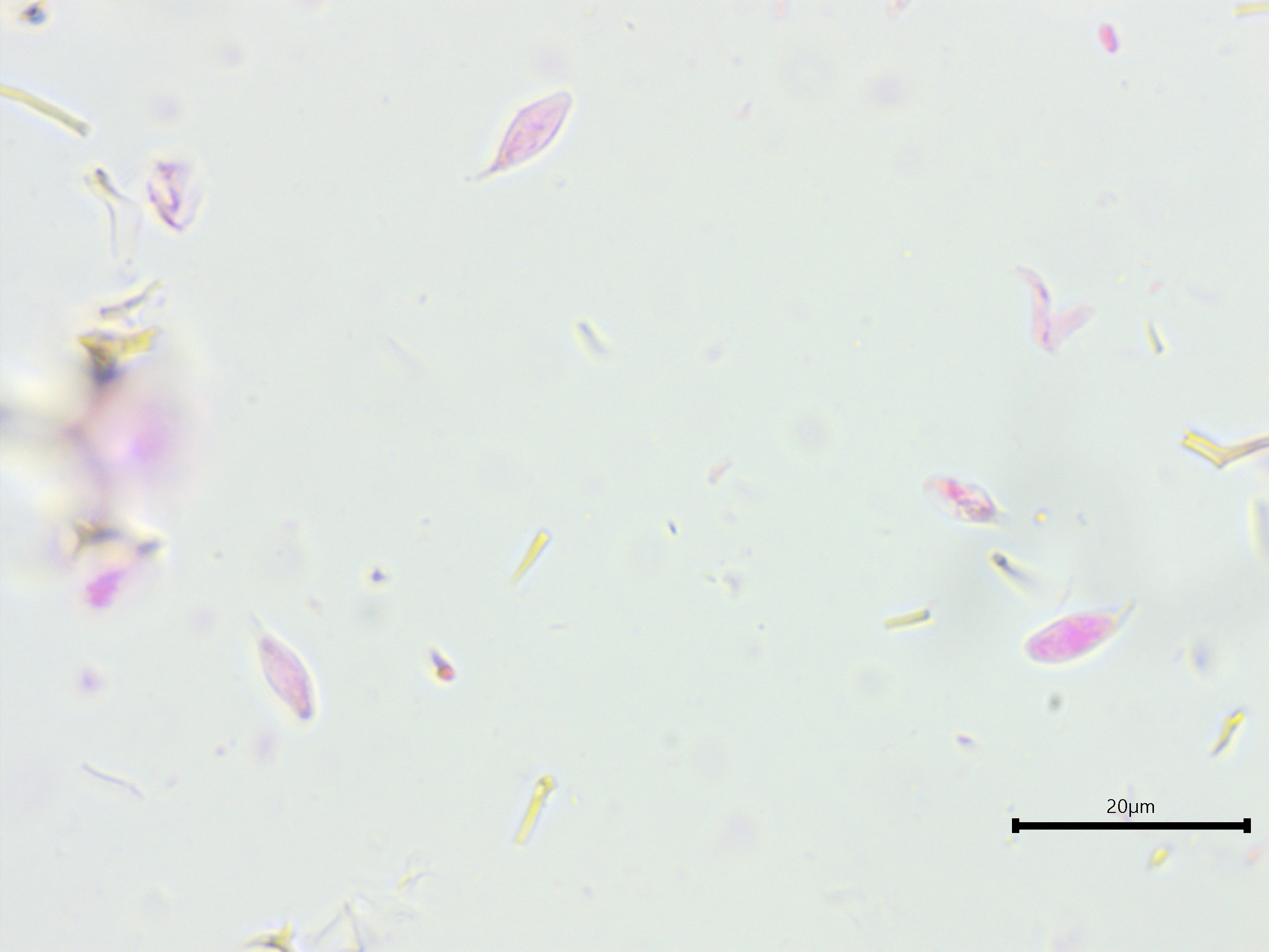
Ellipsoid-fusoid spores.
The extension on the spore tip is amyloid.
Likely clamp connection on a generative hypha.
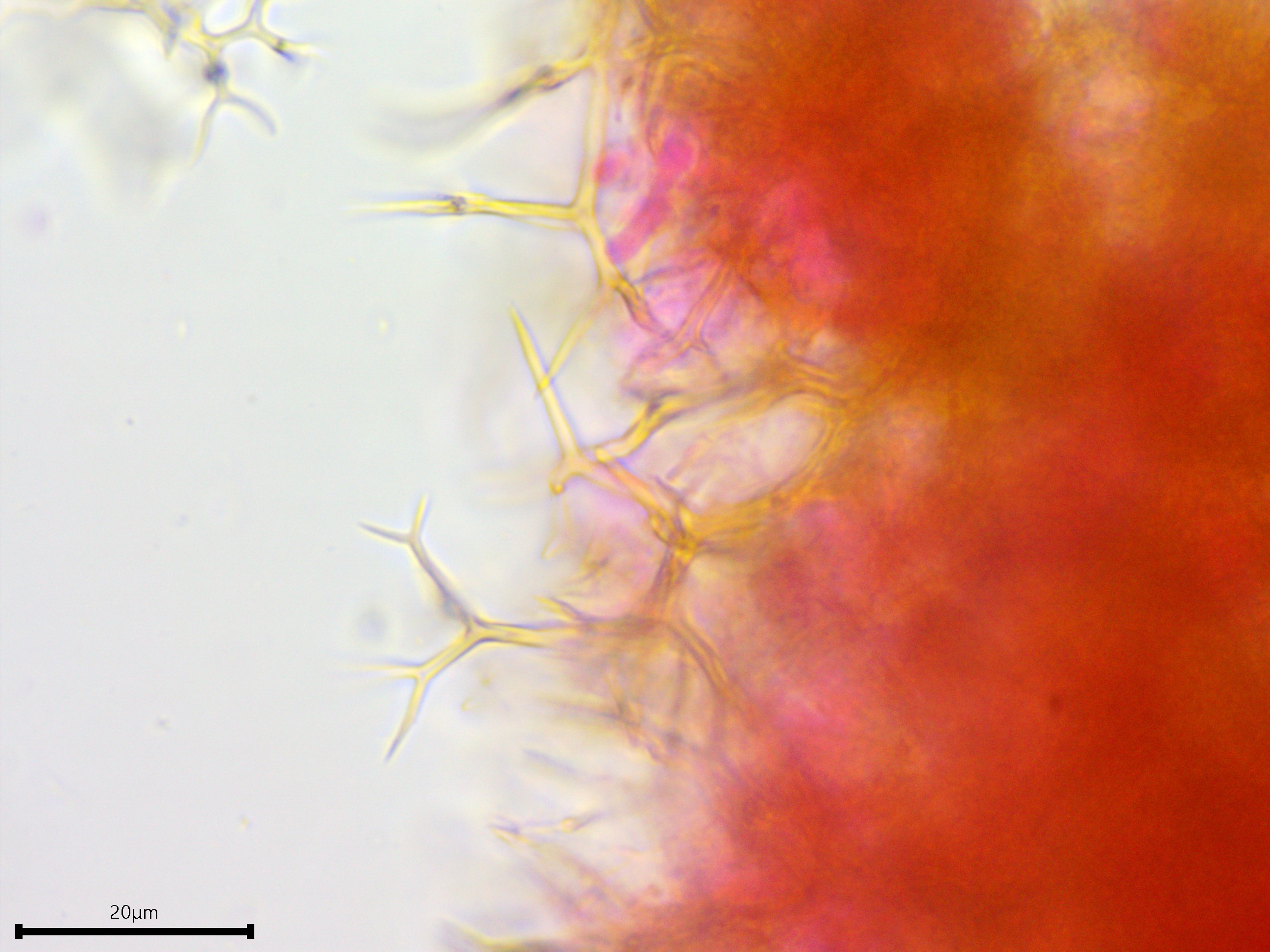
Needle-like dichohyphidia.
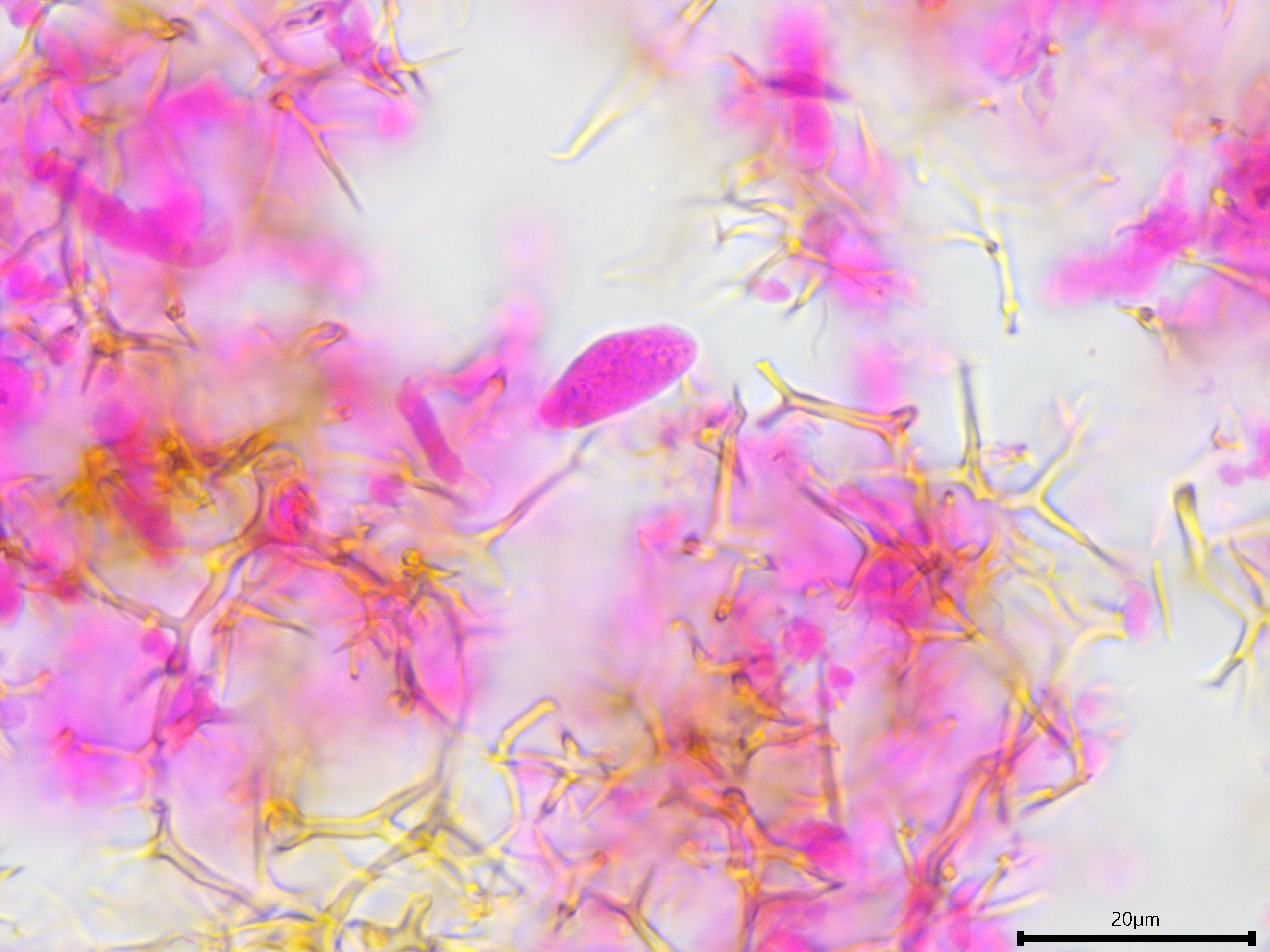
Gloeocystidium.
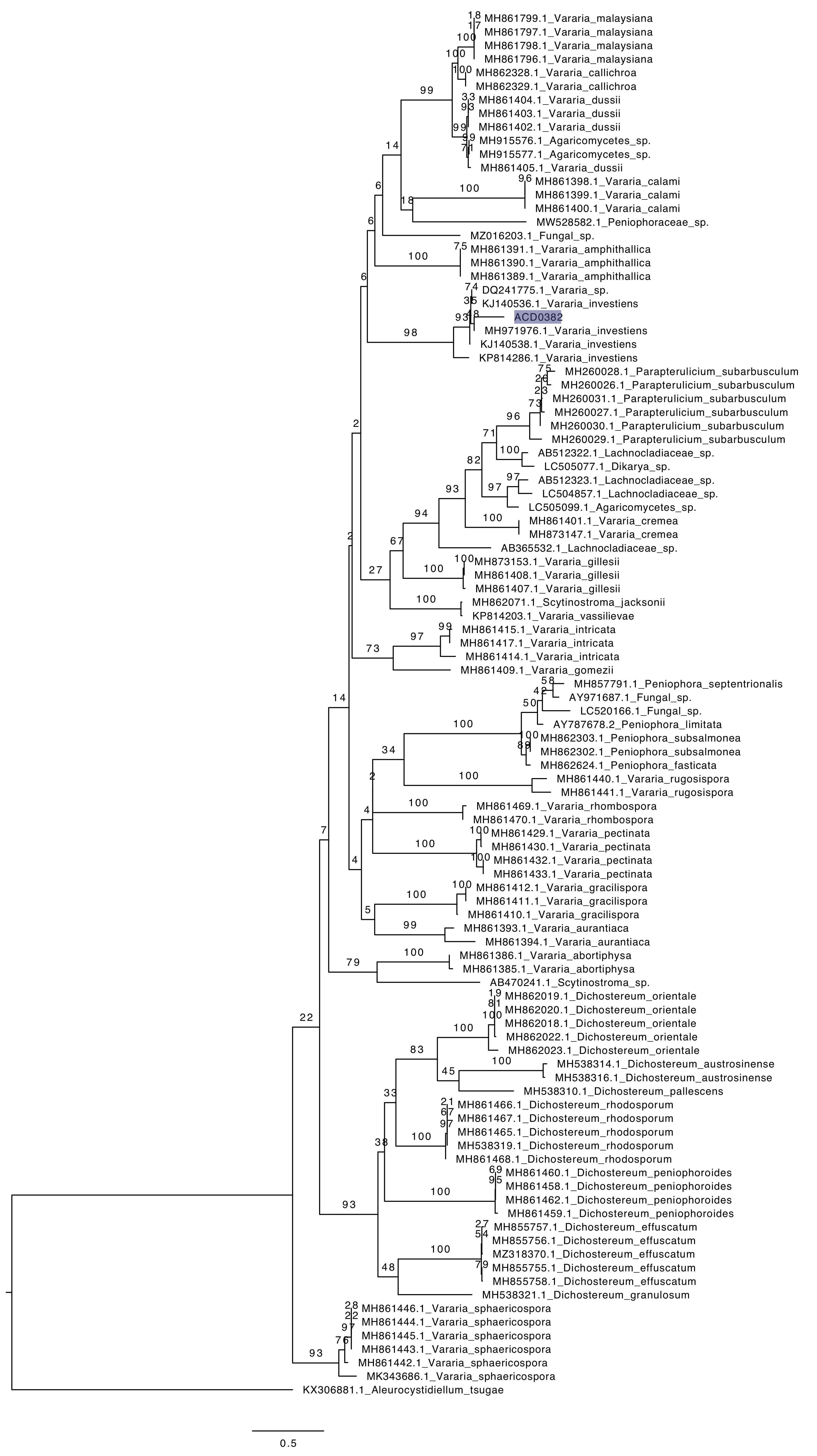
Phylogenetic tree of ITS rDNA sequences from the studied specimen (highlighted) and selected vouchered specimens on GenBank. The sequences were processed with ITSx to remove the flanking SSU and LSU partial sequences, aligned in SeaView with MUSCLE, and made into a tree with RAxML using 100 bootsrap replicates and the GTRGAMMA substitution model. Aleurocystidiellum tsugae serves as the outgroup.