Tubulicrinis accedens (Bourdot & Galzin) Donk
Basidiomycota: Agaricomycotina: Agaricomycetes: Hymenochaetales: Tubulicrinaceae: Tubulicrinis ![]()
![]()
Introduction
There are about 46 species in the genus Tubulicrinis, with Tubulicrinis glebulosus serving as the type species (He et al. 2020). Like far too many crusts out there, Tubulicrinis is sadly understudied beyond the basic taxonomic work that brought the group's species concepts into existence in the first place. That's despite the fact that Tubulicrinis crusts possess distinct sterile structures called lyocystidia whose evolution and ecological function are a complete mystery. Regarding Tubulicrinis accedens, the specific epithet comes from the Latin word accedere meaning "to approach". This fungus was first described as a subspecies, Peniophora glebulosa subsp. accedens, which suggests that accedens refers to an appearance "approaching" that of Peniophora glebulosa (now known as Tubulicrinis glebulosus). Without that context, though, the name doesn't really say much, and even after knowing it, who cares? Let's revise history and say that the "accedens" is a descriptor for how this fungus approaches the heart, filling us with love and reminding us that crusts are undoubtably the best fungi.
Description
Ecology: Presumably saprotrophic, on both deciduous and coniferous wood. The documented specimen was found on the bottom of a fallen, barkless Pinus strobus (white pine) trunk. Widely distributed across temperate North America and Europe according to MyCoPortal records.
Basidioma: Thin, effused, smooth, with a pruinose to arachnoid consistency and abrupt, undifferentiated margin, lilac grey in color.
Chemical reactions: NA
Spore print: White.
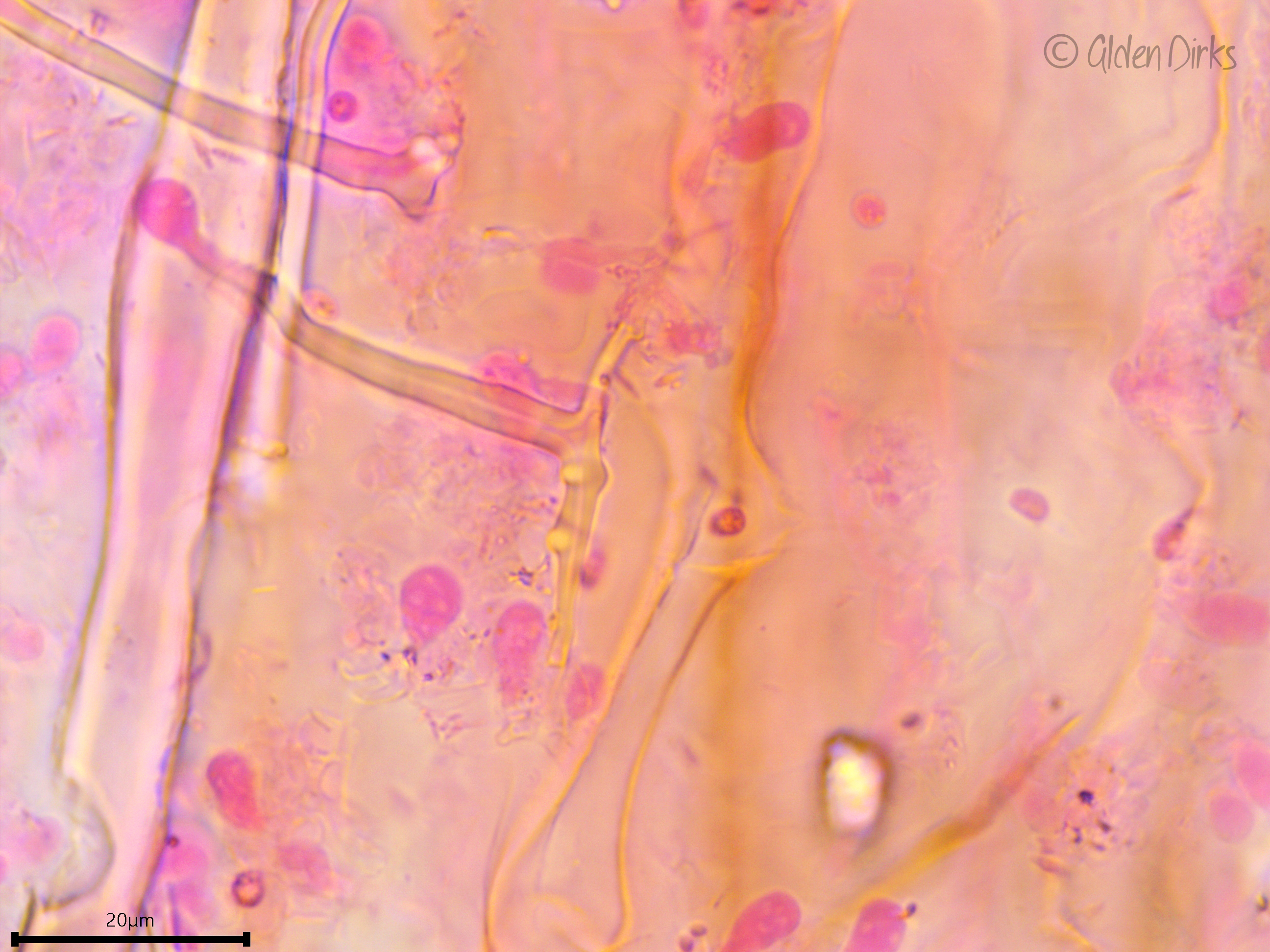
Hyphal system: Monomitic, clamped, 1.5–3 µm wide.
Basidia: Subclavate to conical, small, terminal, with four sterigmata and a basal clamp; length (7.9) 8.8–12.5 (15.2) µm, width (4.2) 4.3–4.9 (5) µm, x̄ = 10.7 ✕ 4.6 µm (n = 10 basidia). This length is likely an underestimate because the basidia were difficult to fully see.
Basidiospores: Ellipsoid to cylindrical, inamyloid, smooth, thin-walled, hyaline; length (3.5) 3.9–4.6 (4.9) µm, width (2.3) 2.6–3 (3) µm, x̄ = 4.2 ✕ 2.8 µm; Q (1.2) 1.4–1.7 (1.8), x̄ = 1.5 (n = 30 basidiospores).
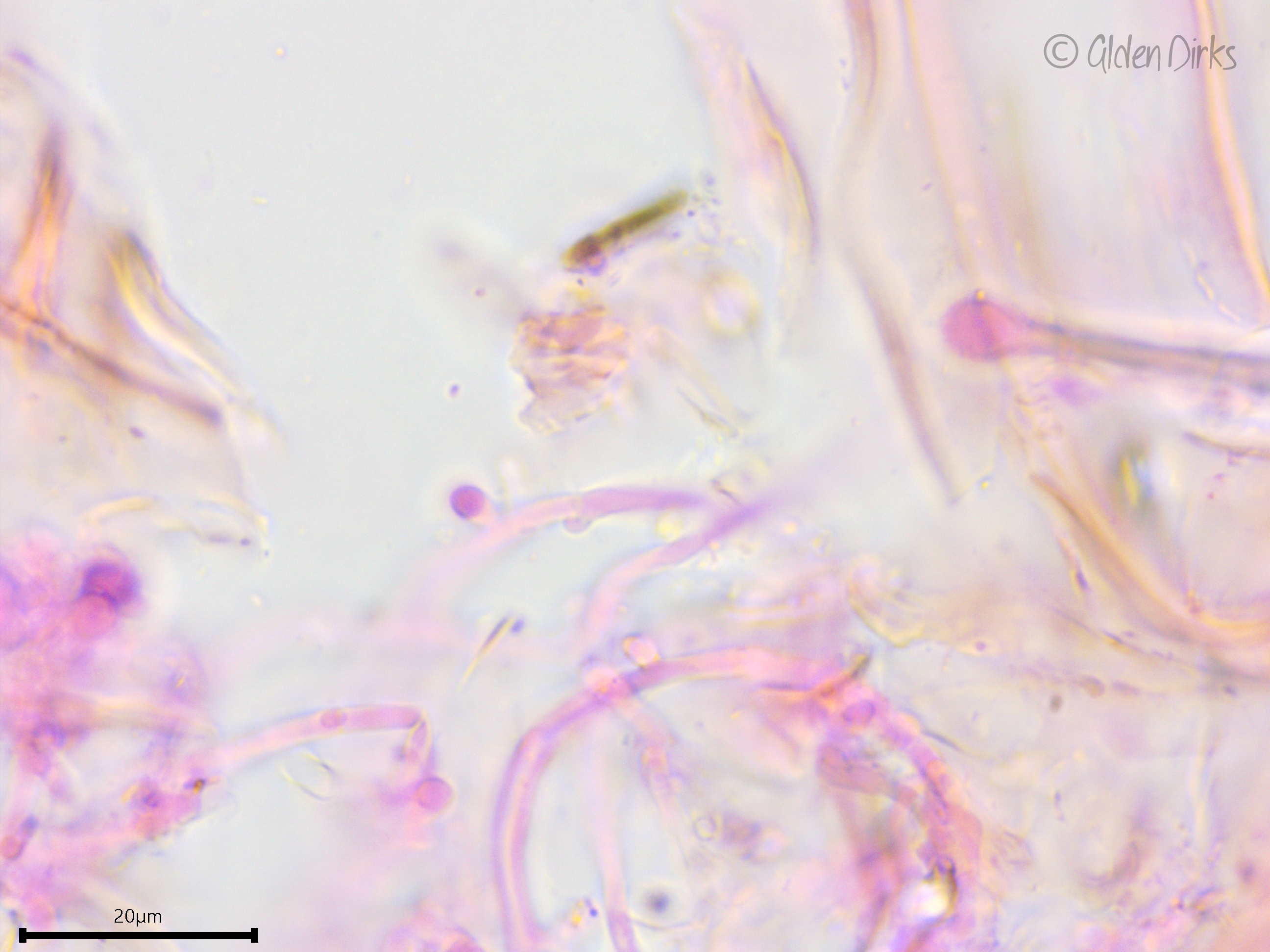
Sterile structures: Lyocystidia very abundant, birooted, consisting of a thick wall that narrows abruptly at the apex to form a globose tip; the globose head, and to a lesser degree the neck, are covered by a somewhat amorphous, chunky, resinous substance that dissolves in KOH; length (37.4) 42.0–50.2 (50.8) µm, basal width (3.2) 3.3–4.0 (4.4) µm, x̄ = 46.1 ✕ 3.7 µm (n = 10 lyocystidia).
Sequences: ITS rDNA (OL756001), LSU rDNA (OL742444).
Notes: All structures measured in 5% KOH stained with phloxine. Bernicchia & Gorjón (2010) report that the lyocystidia of Tubulicrinis accedens turn greyish in Melzer's reagent, but I did not observe this reaction.
Specimens Analyzed
ACD0414, iNat78264072; 10 May 2021; Saginaw Forest, Washtenaw Co., MI, USA, 42.2770 -83.8068; leg. & det. Alden C. Dirks, ref. Bernicchia & Gorjón (2010); University of Michigan Fungarium MICH 352299 .
References
He, X., Shi, Z.-J., & Zhao, C.-L. (2020). Morphological and molecular identification of two new species of Tubulicrinis (Hymenochaetaceae, Hymenochaetales) from southern China. Mycoscience, 61(4), 184–189.
Links
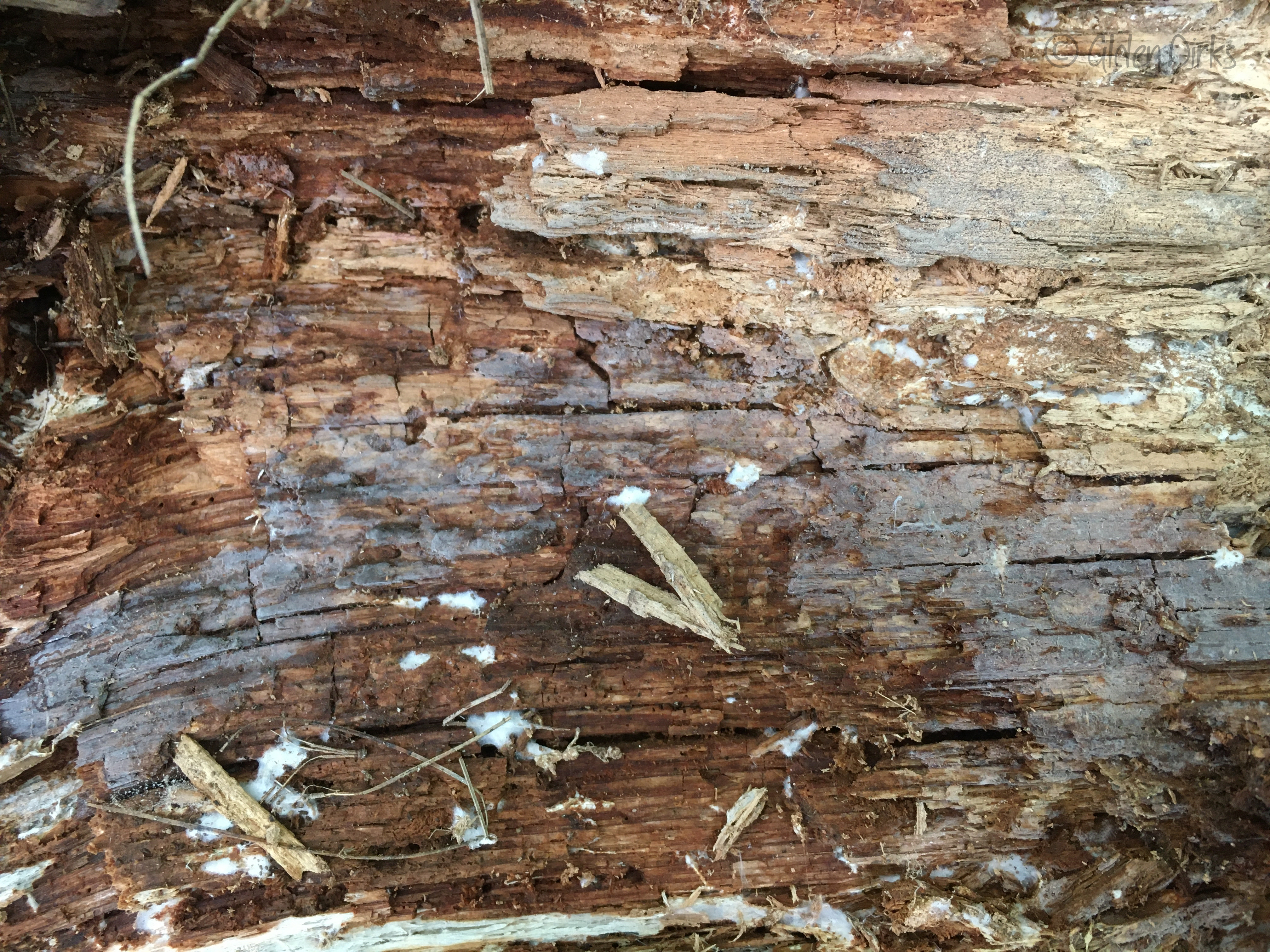
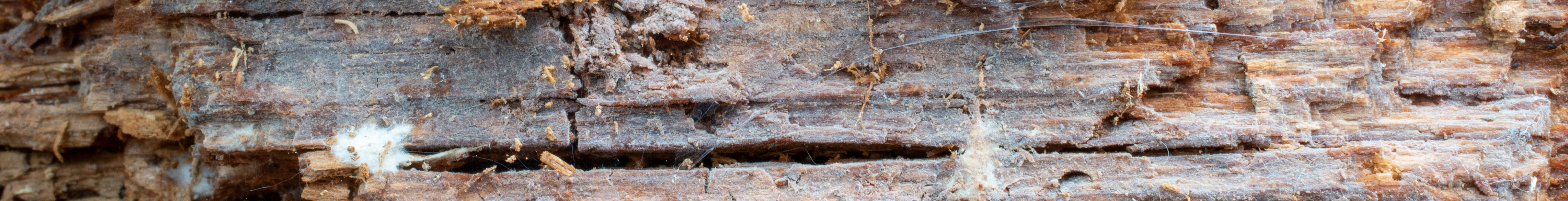
Tubulicrinis accedens constellation on the bottom of a fallen white pine trunk.
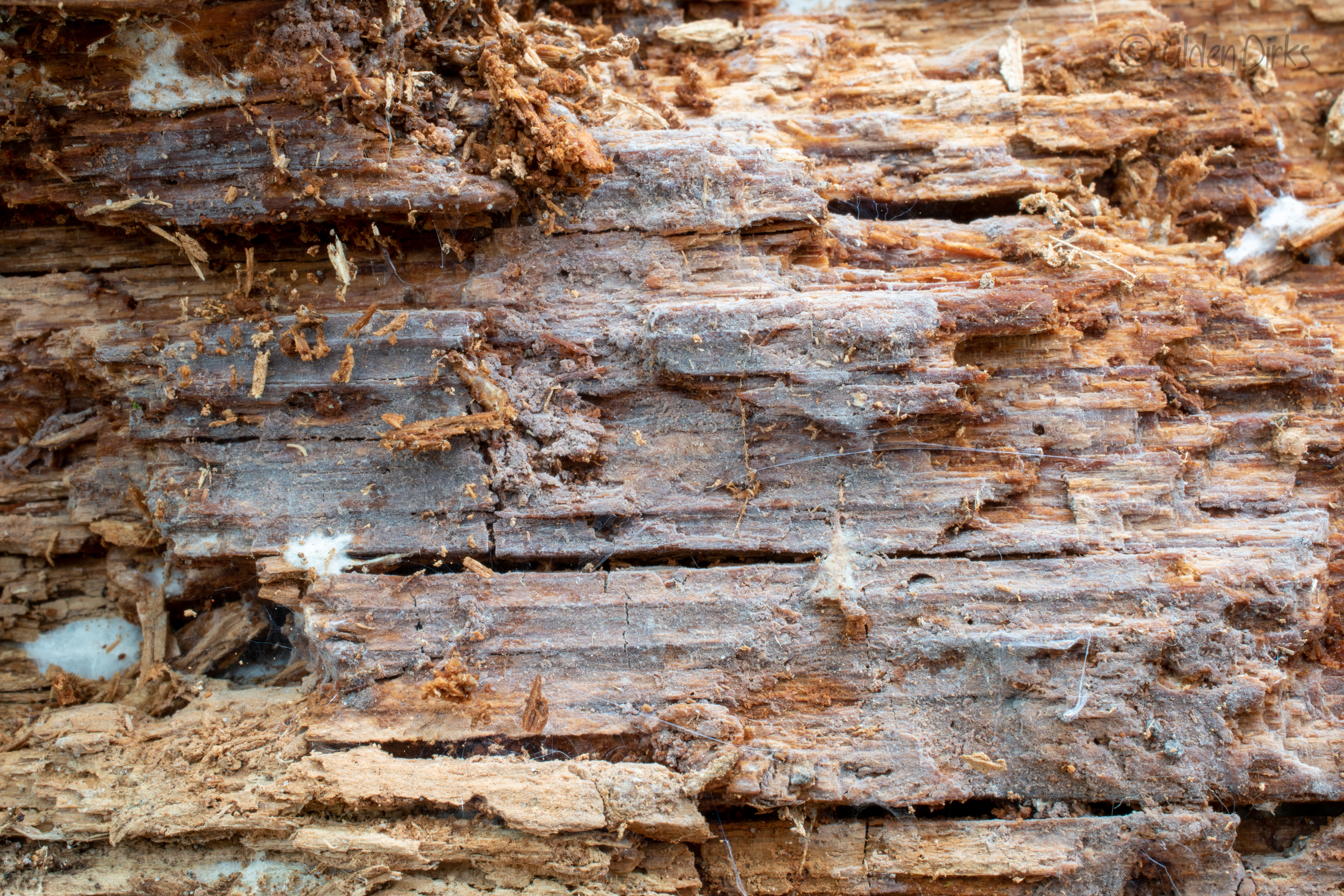
Close-up image of the Tubulicrinis accedens basidioma.
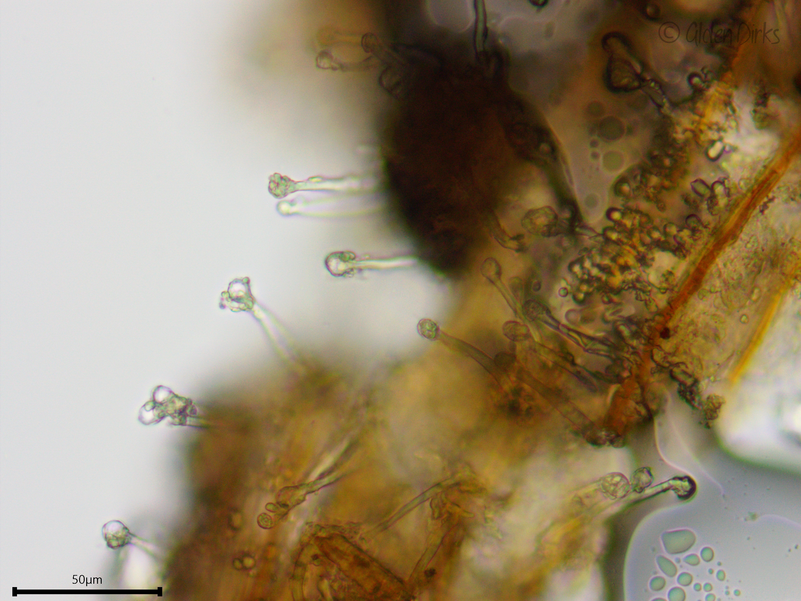
Abundant lyocystidia (water).
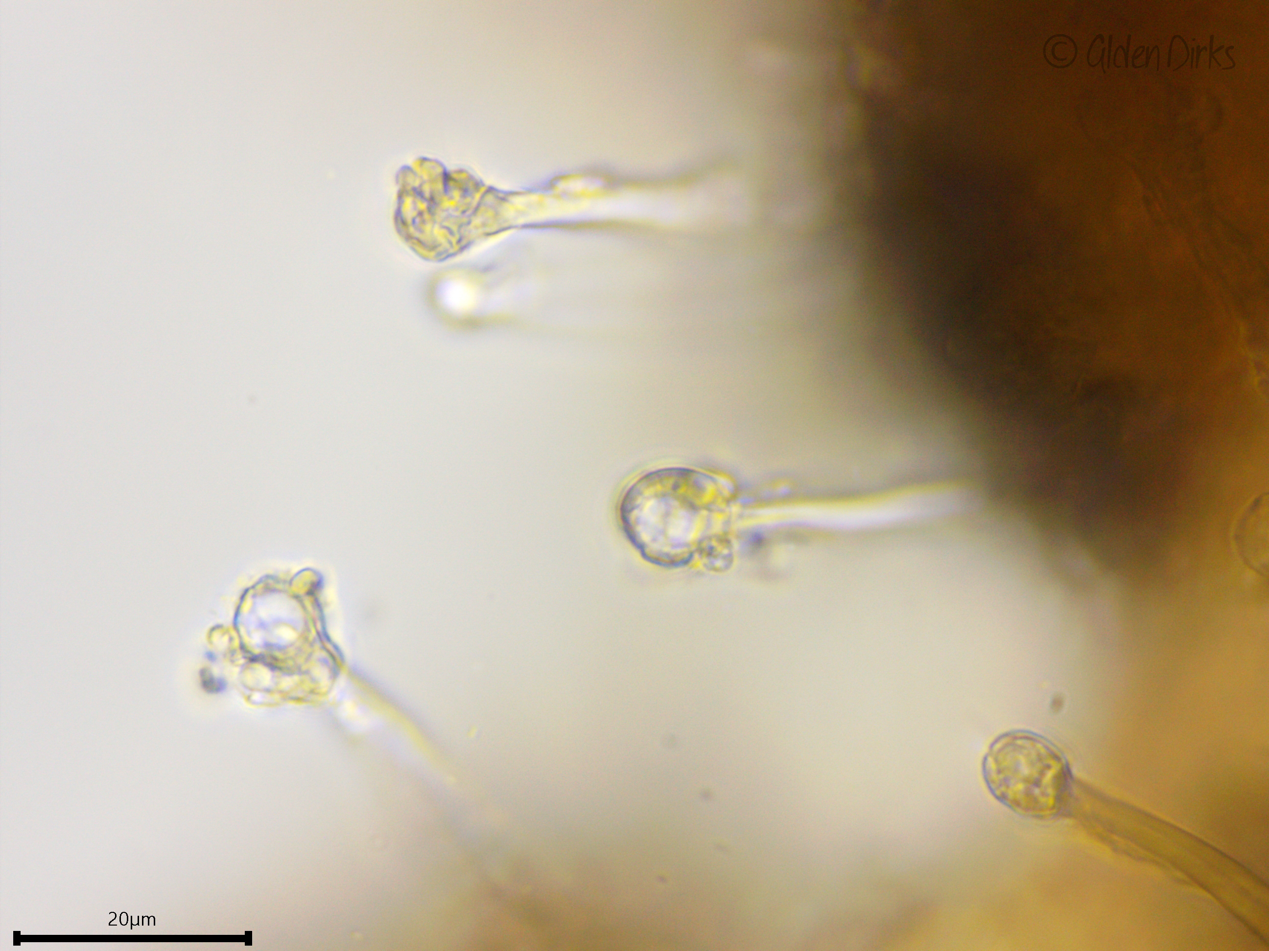
Closer look at the resinous material coating the lyocystidia heads (visible in water, dissolving in KOH).
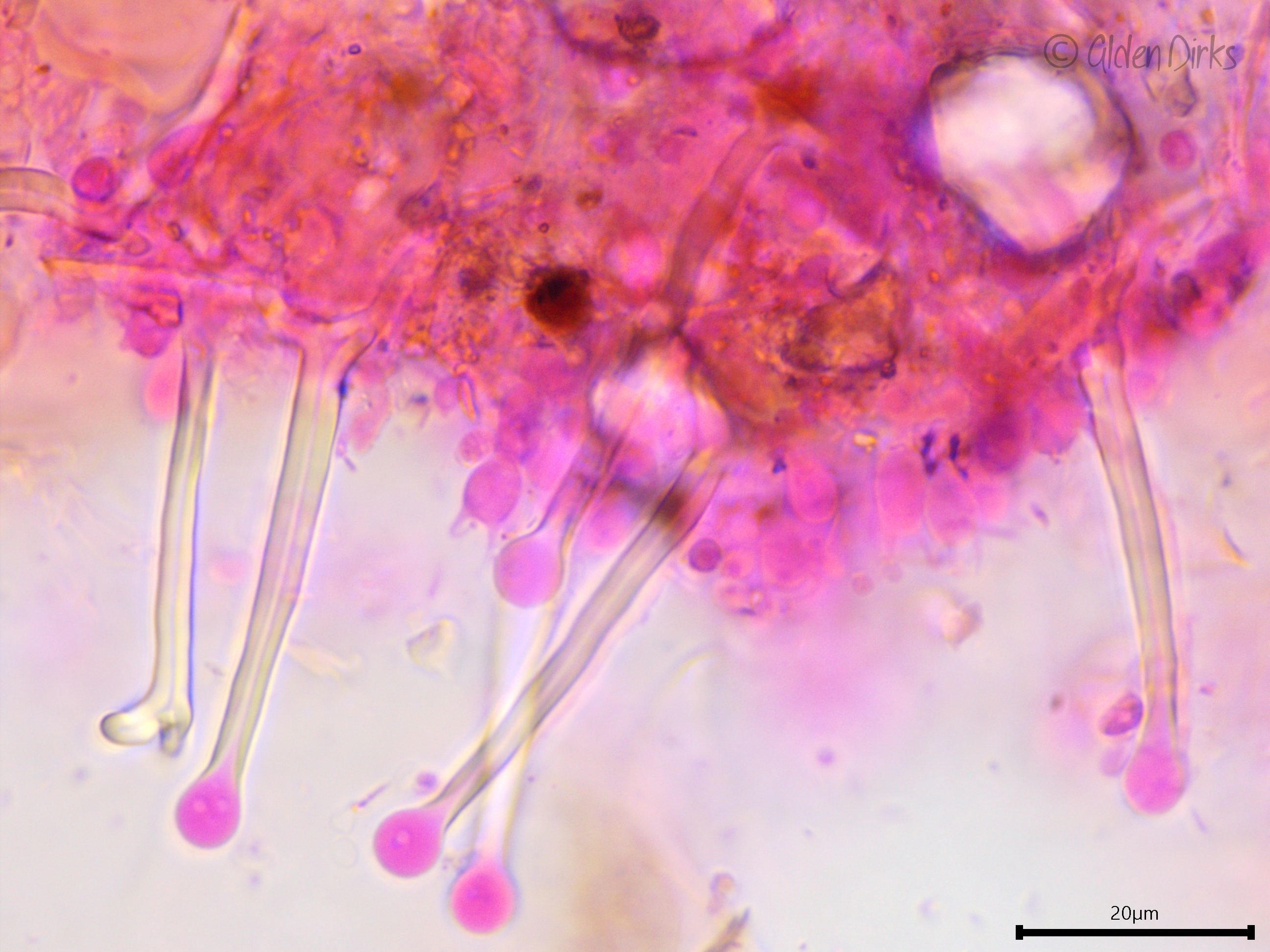
Lyocystidia in KOH stained with phloxine, showing the very thick walls that abruptly narrow at the tip to form a capitate head.
Birooted lyocystidium.
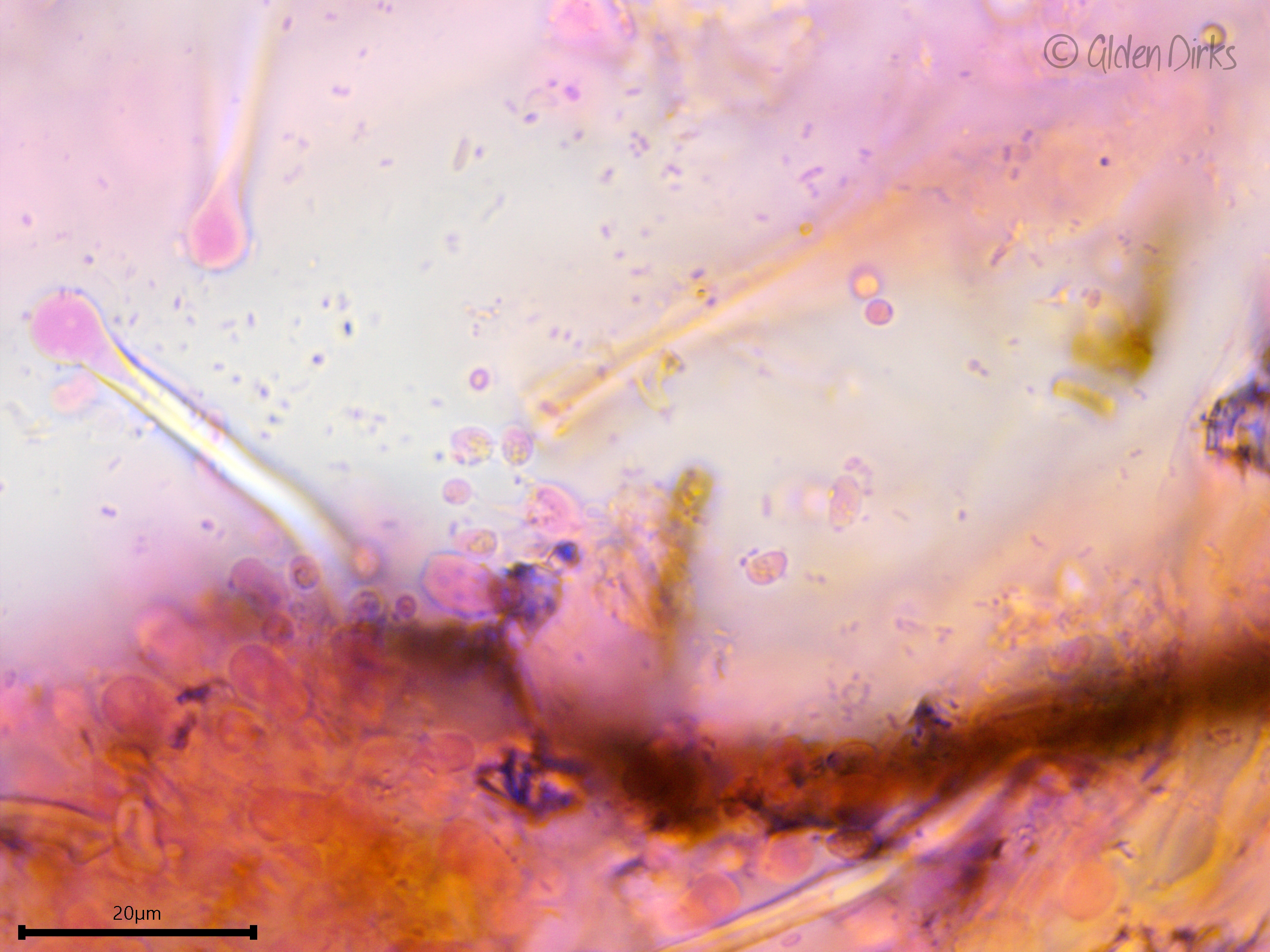
Basidium with four sterigmata.
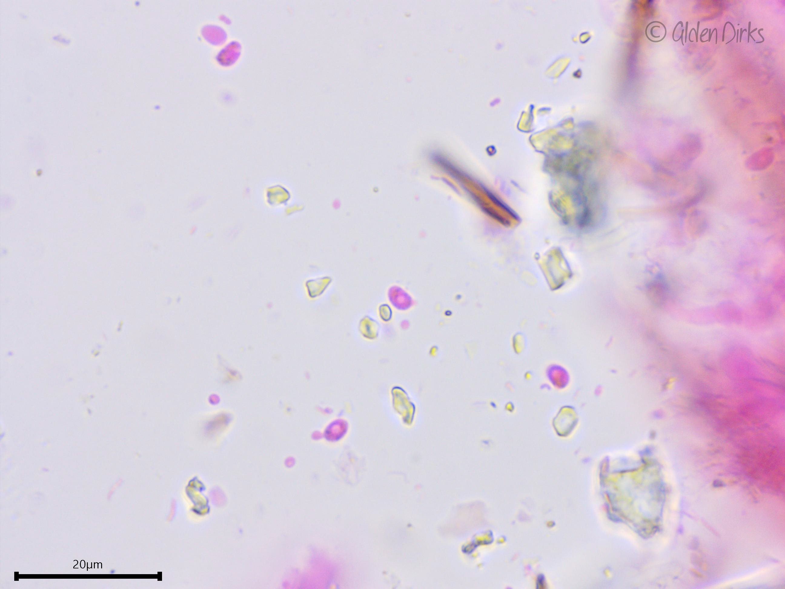
Small basidiospores.
Clamp connection.
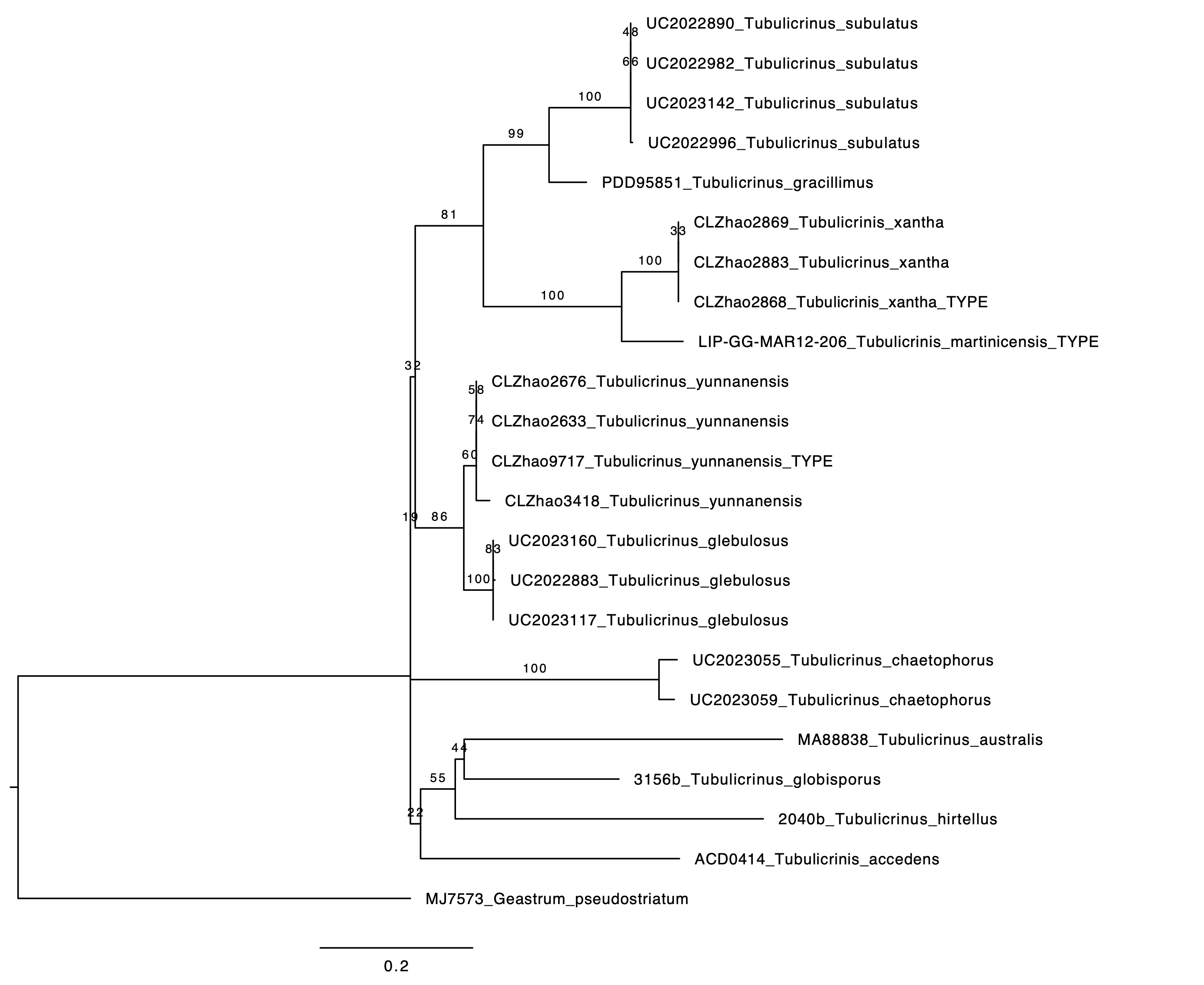
Phylogenetic tree of ITS rDNA sequences from the studied specimen and selected vouchered specimens on GenBank, modeled after that of He et al. (2020). The sequences were processed with ITSx to remove the flanking SSU and LSU partial sequences, aligned in SeaView with MUSCLE, and made into a tree with RAxML using 100 bootsrap replicates and the GTRGAMMA substitution model. Geastrum pseudostriatum serves as the outgroup. There are too few LSU sequences to make a meaningful concatenated tree.