Punctularia strigosozonata (Schwein.) P.H.B. Talbot
Tree Bacon
Basidiomycota: Agaricomycotina: Agaricomycetes: Corticiales: Punctulariaceae: Punctularia ![]()
![]()
Introduction
To my knowledge, nobody has tried eating tree bacon, but when you peel a line of these mushrooms off a log, the strangely flappy, rubbery fruiting bodies seem like they'd fit right in on a sizzling hot pan. I am unsure how common Punctularia strigosozonata is in North America, but this was my first time finding this distinct crust, and it is reported as rare in Europe (Bondartseva et al. 2000). The wrinkled hymenophore and gelatinous to ceraceous consistency are reminiscent of Phlebia species in Meruliaceae (Polyporales), but Punctularia strigosozonata actually belongs in Corticiales, in the family Punctulariaceae, which only contains two other genera.
As a white-rot fungus, Punctularia strigosozonata has received research attention for its powerful lignin-degrading enzymes. To better understand the evolution of wood decay, mycologists analyzed 31 genomes, including that of Punctularia strigosozonata, and found that the common ancestor of Agaricomycetes was probably a white-rot species (Floudas et al. 2012). In this same study, it was estimated that lignin-degrading enzymes evolved around 300 million years ago. This timing supports the hypothesis that the evolution of lignin in plants was what caused massive coal deposits to form in the Carboniferous period and the evolution of lignin degradation by white-rot Agaricomycetes some time later was what subsequently ended the era of coal.
Description
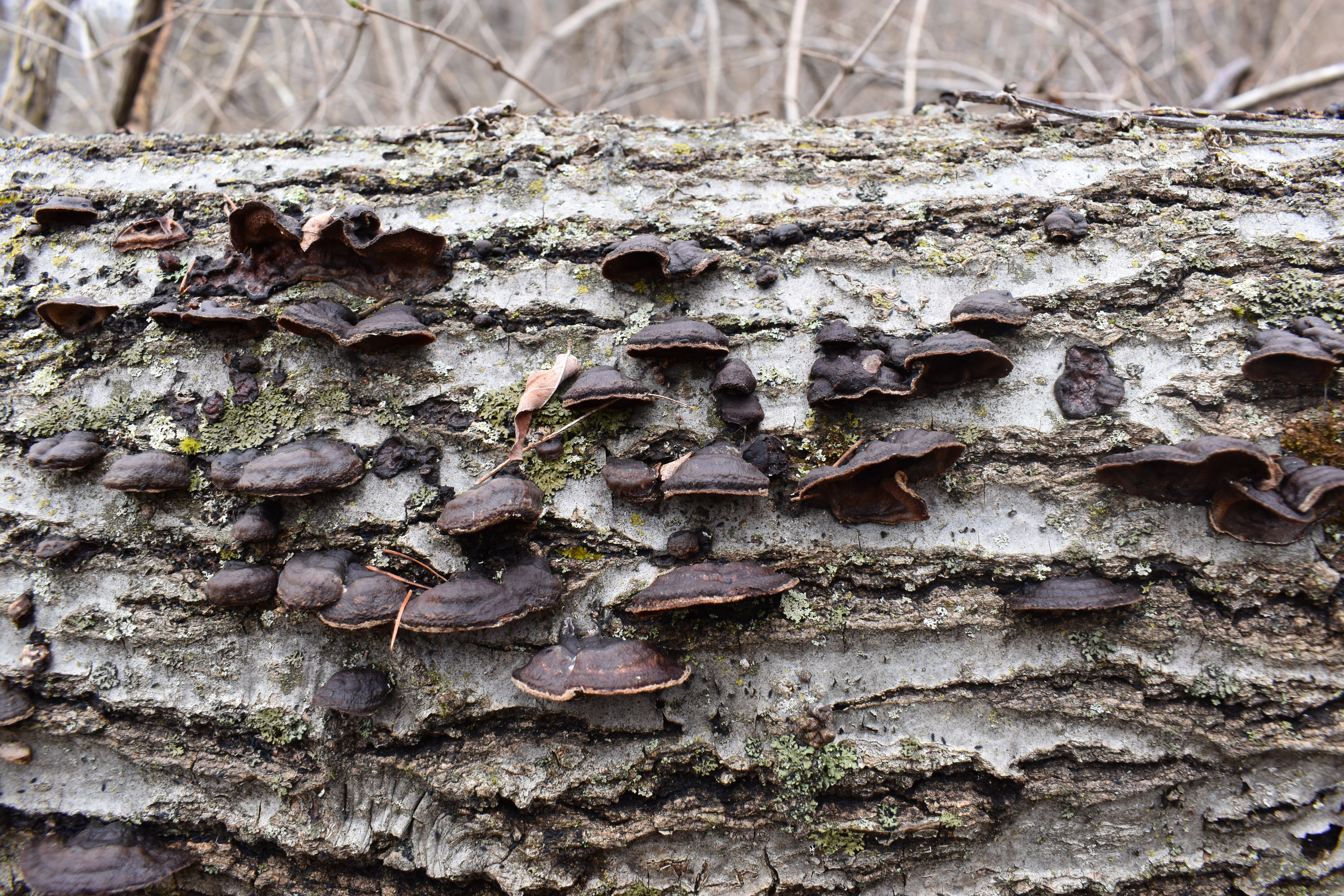
Ecology: Apparently globally distributed, preferring hardwood logs, especially Populus spp., but also reported on gymnosperms (Crusts and Jells).
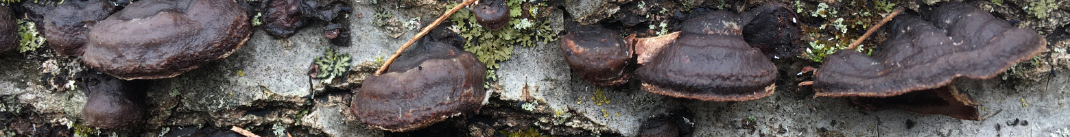
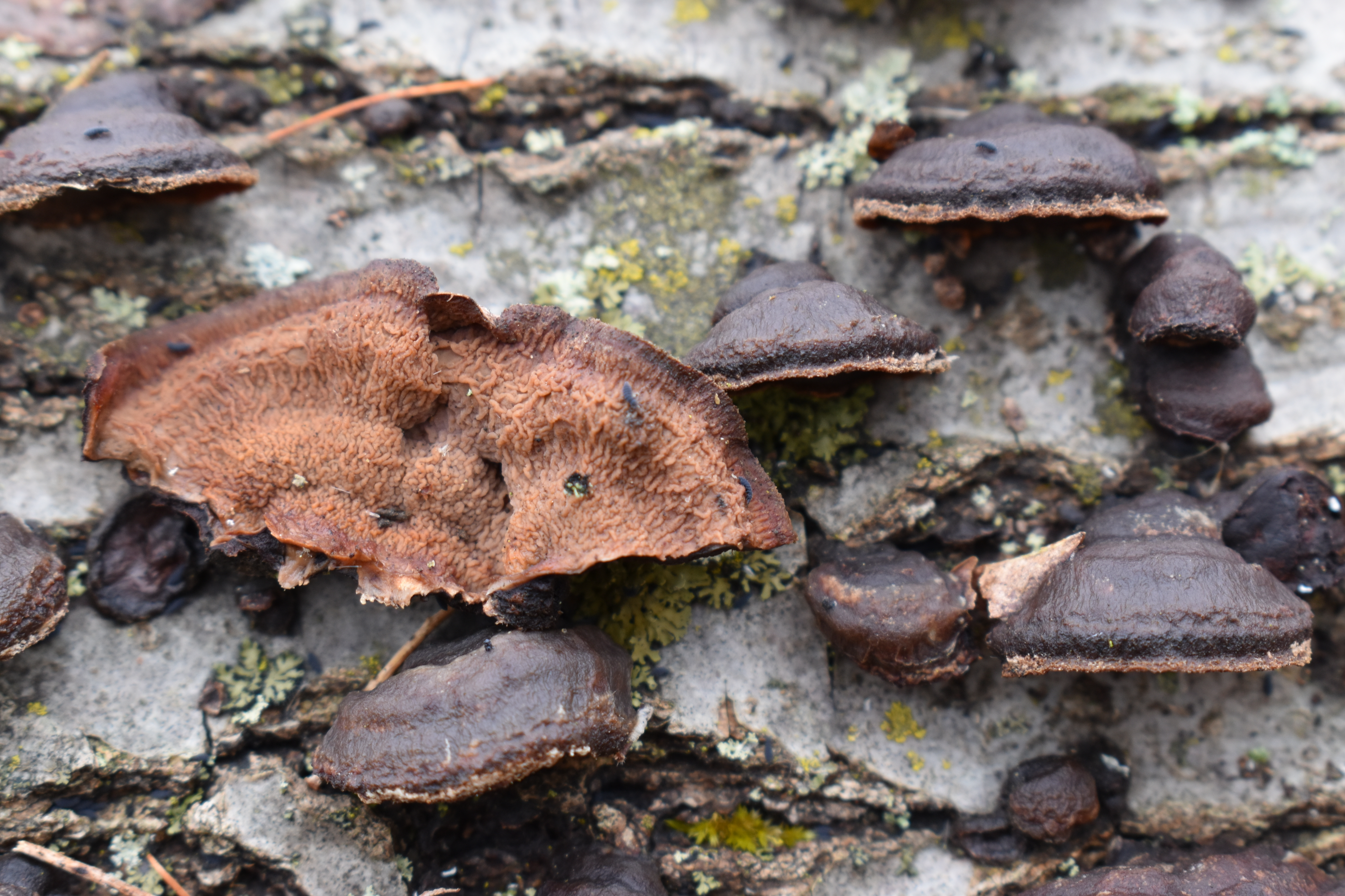
Basidiocarp: Variable, ranging from completely effused, more typically effused-reflexed, sometimes dimidiate; cap surface dark reddish brown, zonate, with concentric ridges of growth and a pale brown margin; hymenophore smooth to phlebioid or merulioid, also described as tuberculate and radially ridged (Bernicchia & Gorjón 2010), orangish brown to maroon or vinaceous; consistency gelatinous or ceraceous to coriaceous, with the look of rendered fat, lending this species its common name "tree bacon"; when dry, cap is tannish and hymenium is light grey to black.
Chemical reactions: NA
Spore print: Whitish, pale cream.
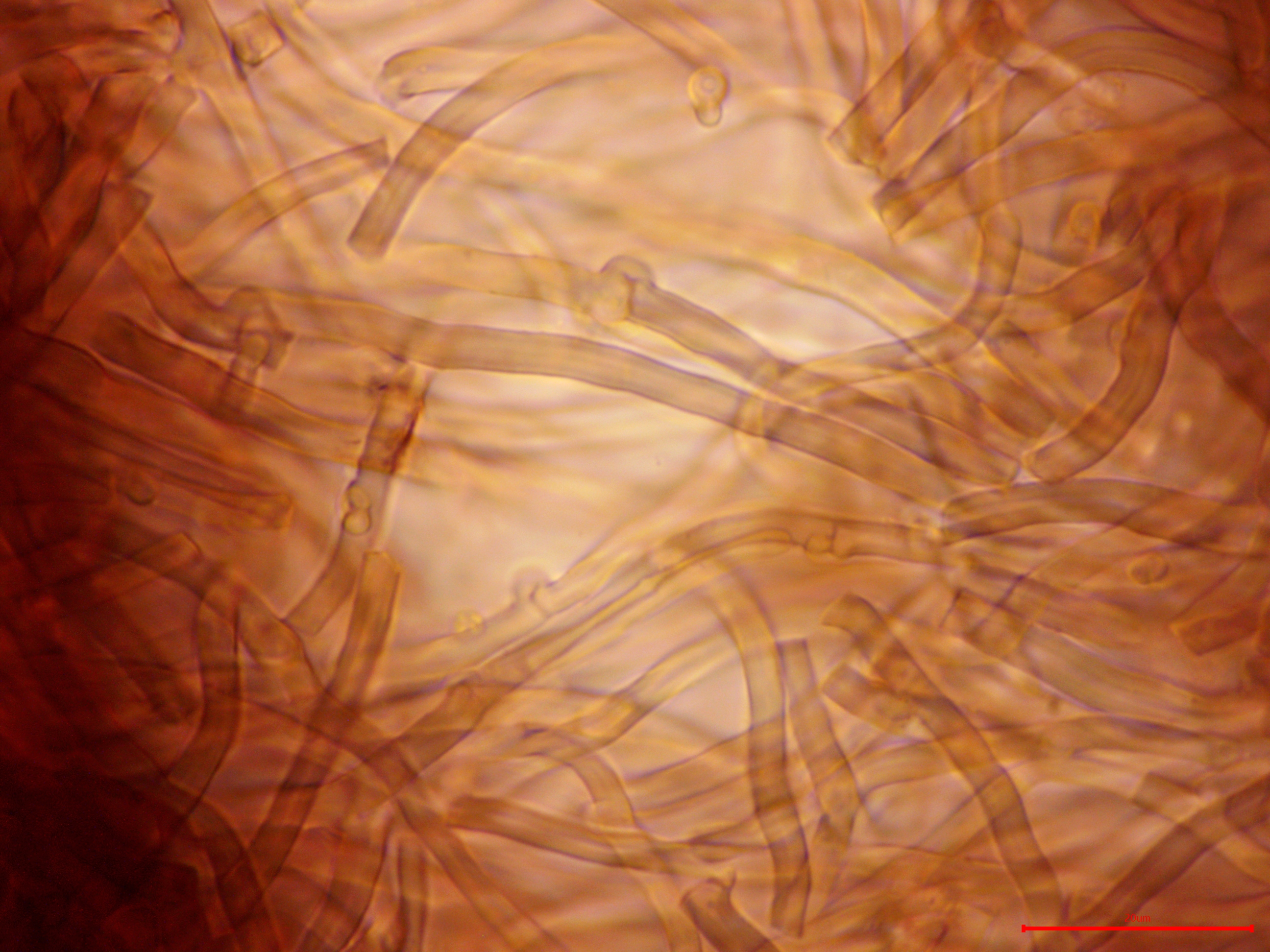
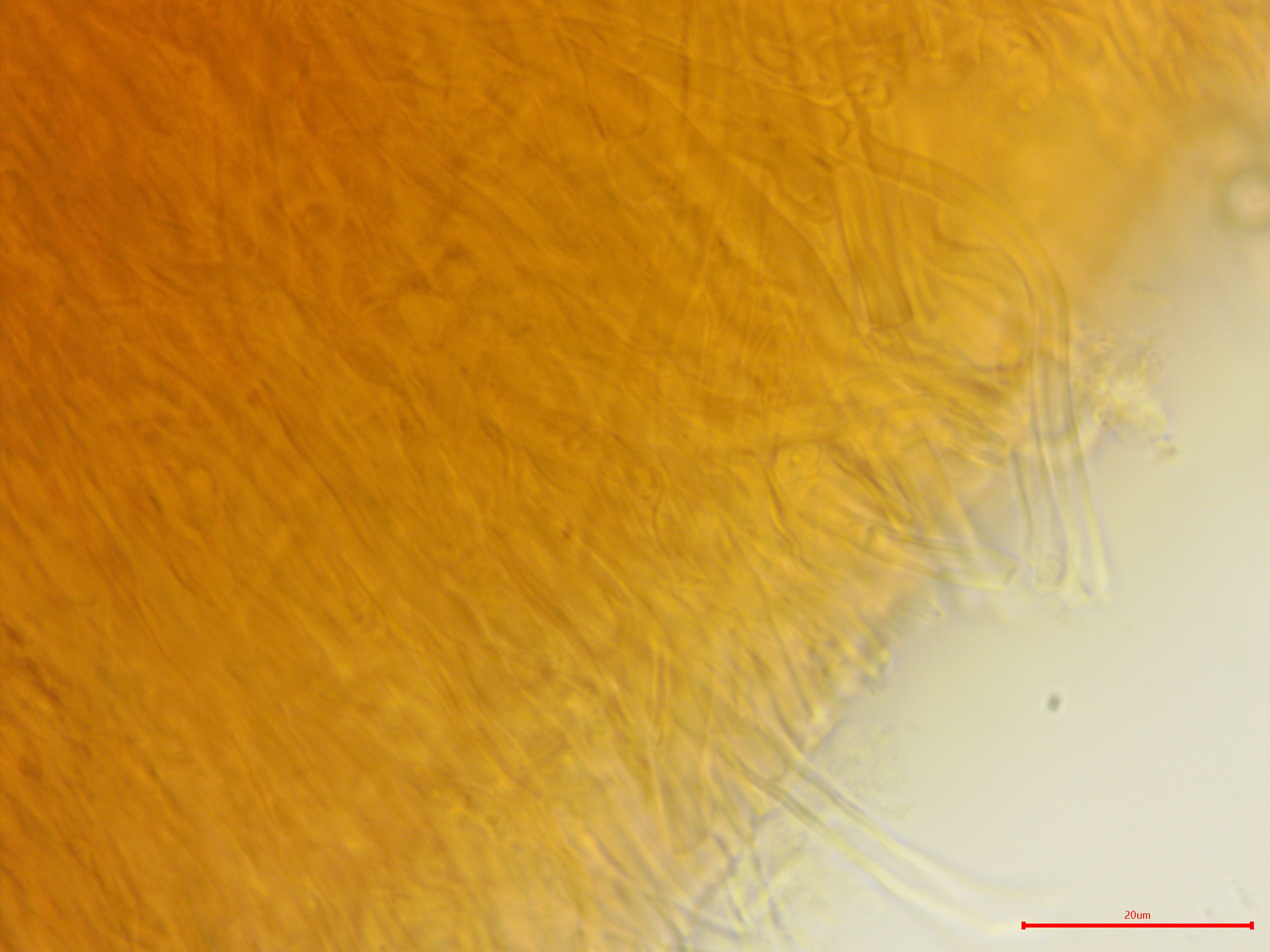
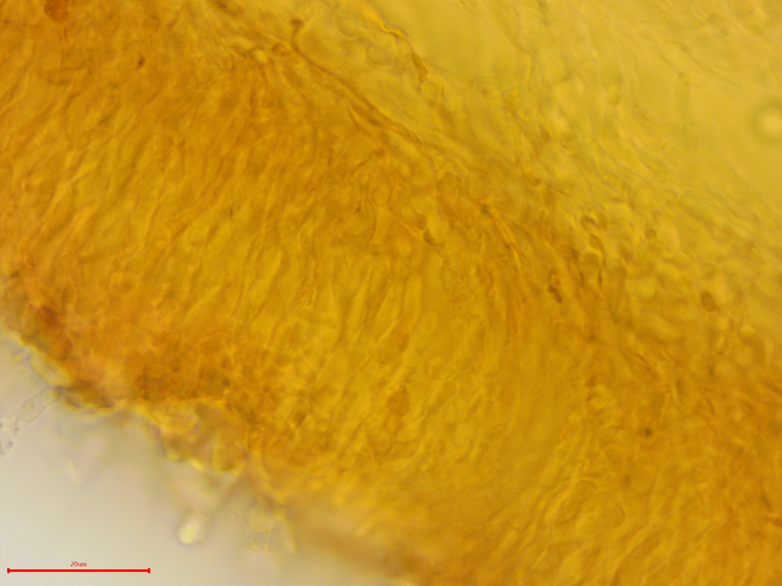
Hyphal system: Monomitic, all septa with clamps; large granules embedded within trama, subhymenium, and hymenium; abhymenial hyphae loose, thick-walled, width (2.7) 3.3–4.4 (4.5) µm, x̄ = 3.8 µm; hyphae in trama thick-walled, resembling skeletohyphae but regularly septate, width (3.3) 3.6–4.6 (4.8) µm, x̄ = 4.1 µm; subhymenial hyphae running parallel to hymenium surface, tightly packed, thin-walled to slightly thick-walled, width (2.5) 2.5–3.6 (4.4) µm, x̄ = 3.0 µm; 10 hyphae per section measured in Melzer's reagent.
Basidia: Clavate, sometimes stalked, with four sterigmata; length (12.6) 13.6–18.3 (19.6) µm, width (4.3) 4.4–5.8 (6.3) µm, x̄ = 16.0 ✕ 5.1 µm (n = 10 basidia measured in Melzer's reagent or KOH stained with phloxine). However, base of basidia not observed and Bernicchia & Gorjón (2010) report length as 40–60 (80) µm.
Basidiospores: Cylindrical, hyaline to yellowish, inamyloid; length (5.4) 6.0–6.9 (7.3) µm, width (3.5) 3.6–4.0 (4.3) µm, x̄ = 6.5 ✕ 3.8 µm; Q (1.5) 1.6-1.8 (1.9), x̄ = 1.7 (n = 30 basidiospores measured in tap water).
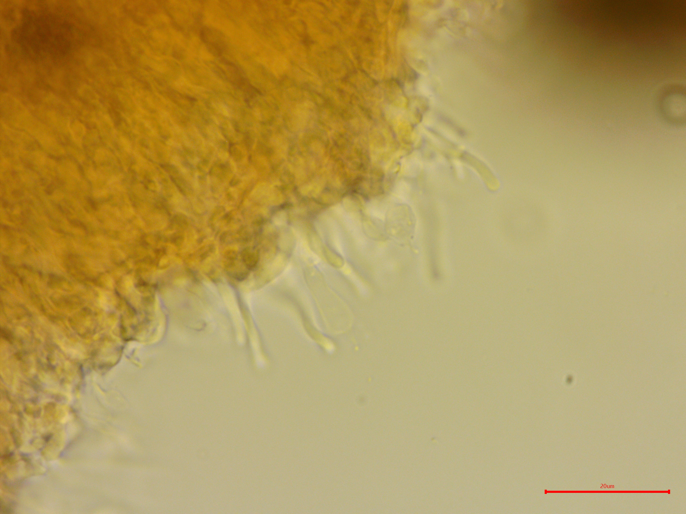
Sterile structures: Dendrohyphidia.
Sequences: NA
Notes: Microscopic features are difficult to observe in this species due to the dense, gelatinous nature of the hyphae. However, this species is easily identified by its macroscopic features.
Specimens Analyzed
ACD0243, iNat40928813; 27 March 2020; Ott Biological Preserve, Calhoun Co., MI, USA, 42.3165 -895.1245; leg. Alden Dirks, det. Alden Dirks, recognized by sight; University of Michigan Fungarium MICH 352230.
References
Bernicchia, A. & Gorjón, S. P. (2010). Fungi Europaei, Volume 12: Corticiaceae s.l. Edizioni Candusso, Italia.
Bondartseva, M. A., Lositskaya, V. M., & Zmitrovich, I. V. (2000). Punctularia strigosozonata (Punctulariaceae) in Europe. Karstenia, 40(1–2), 9–10. DOI
Floudas, D., Binder, M., Riley, R., Barry, K., Blachette, R. A., Henrissat, B., … Hibbett, D. S. (2012). The Palaeozoic origin of enzymatic lignin decomposition reconstructed from 31 fungal genomes. Science, 336(June), 1715–1719.
Links
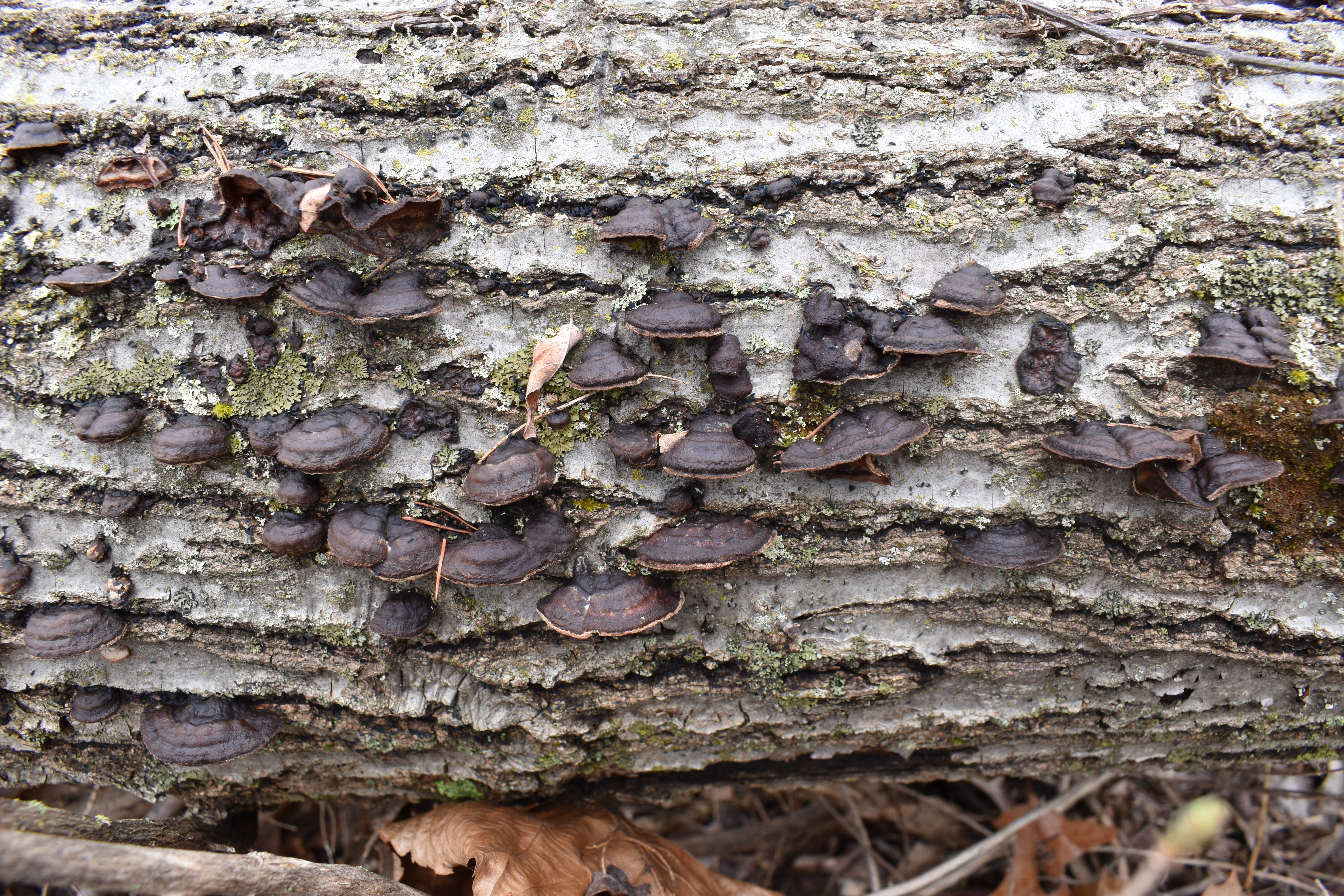
Effused-reflexed to dimidiate fruiting bodies of Punctularia strigosozonata.
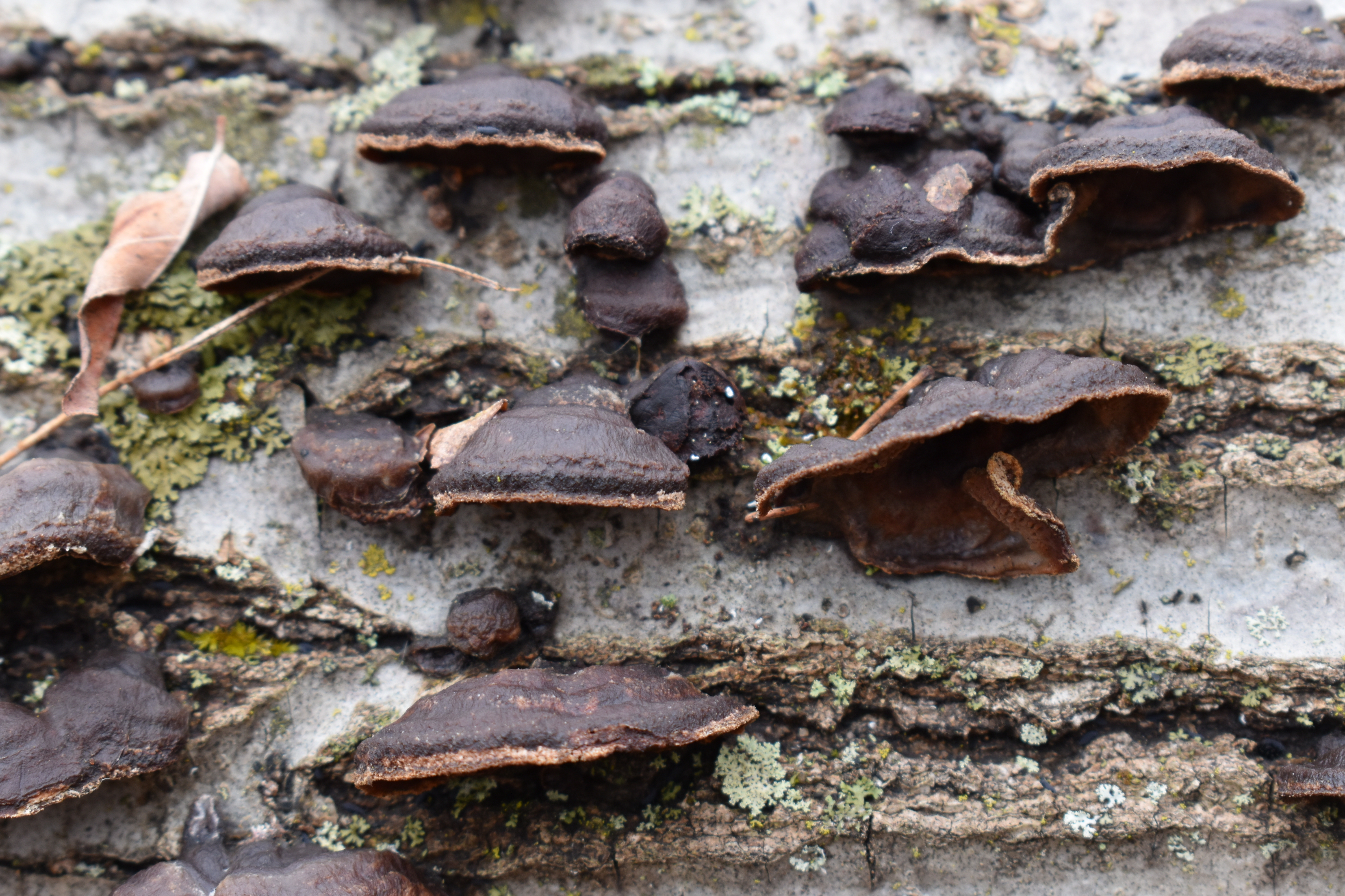
The cap margin is a different color than the rest of the cap.
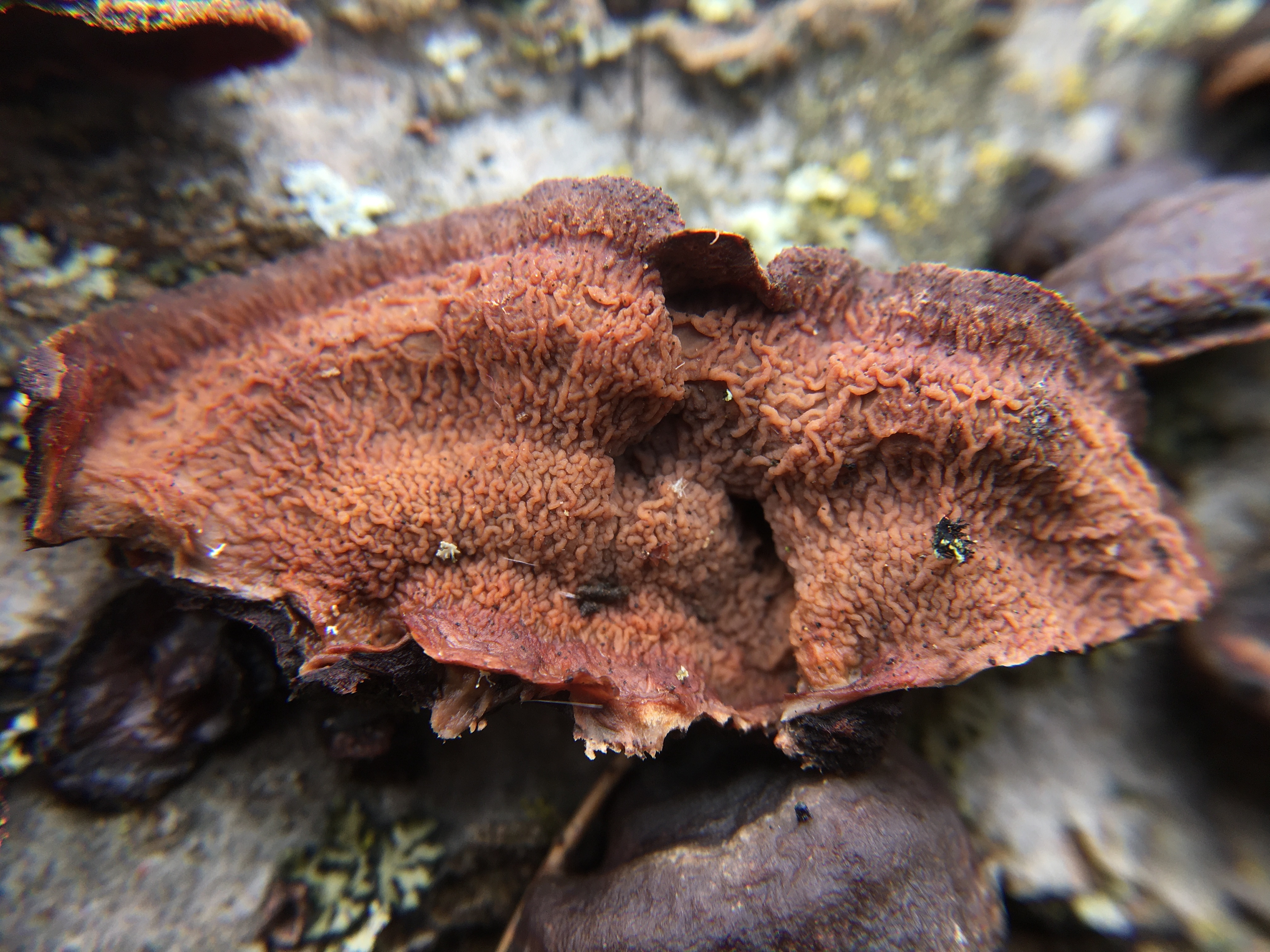
The hymenophore is radially wrinkled.
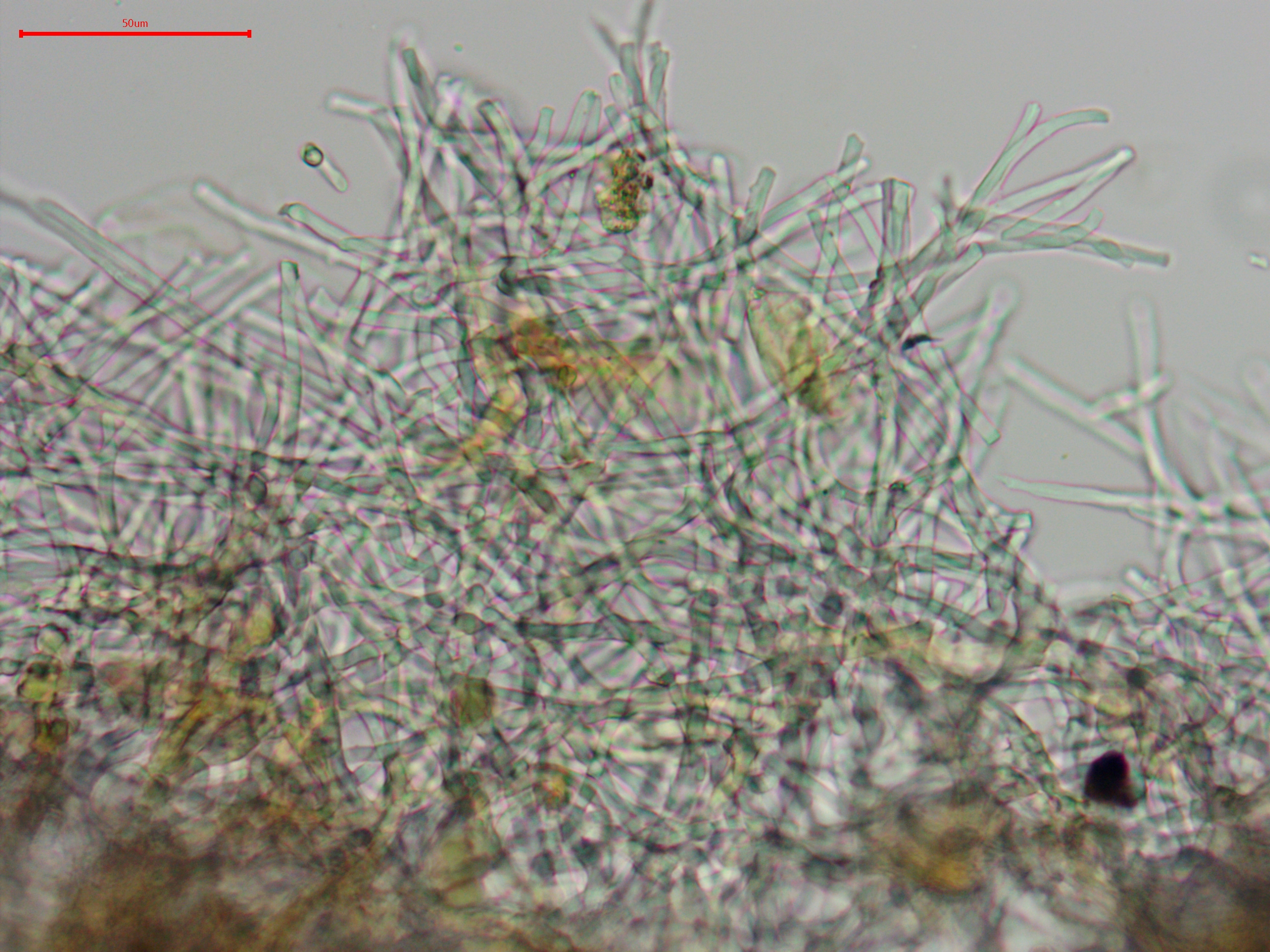
Abhymenial surface hyphae.
Brown subabhymenial surface hyphae.
Thick-walled hyphae in the trama.
Subhymenial hyphae running into the hymenium.
Clavate basidium.
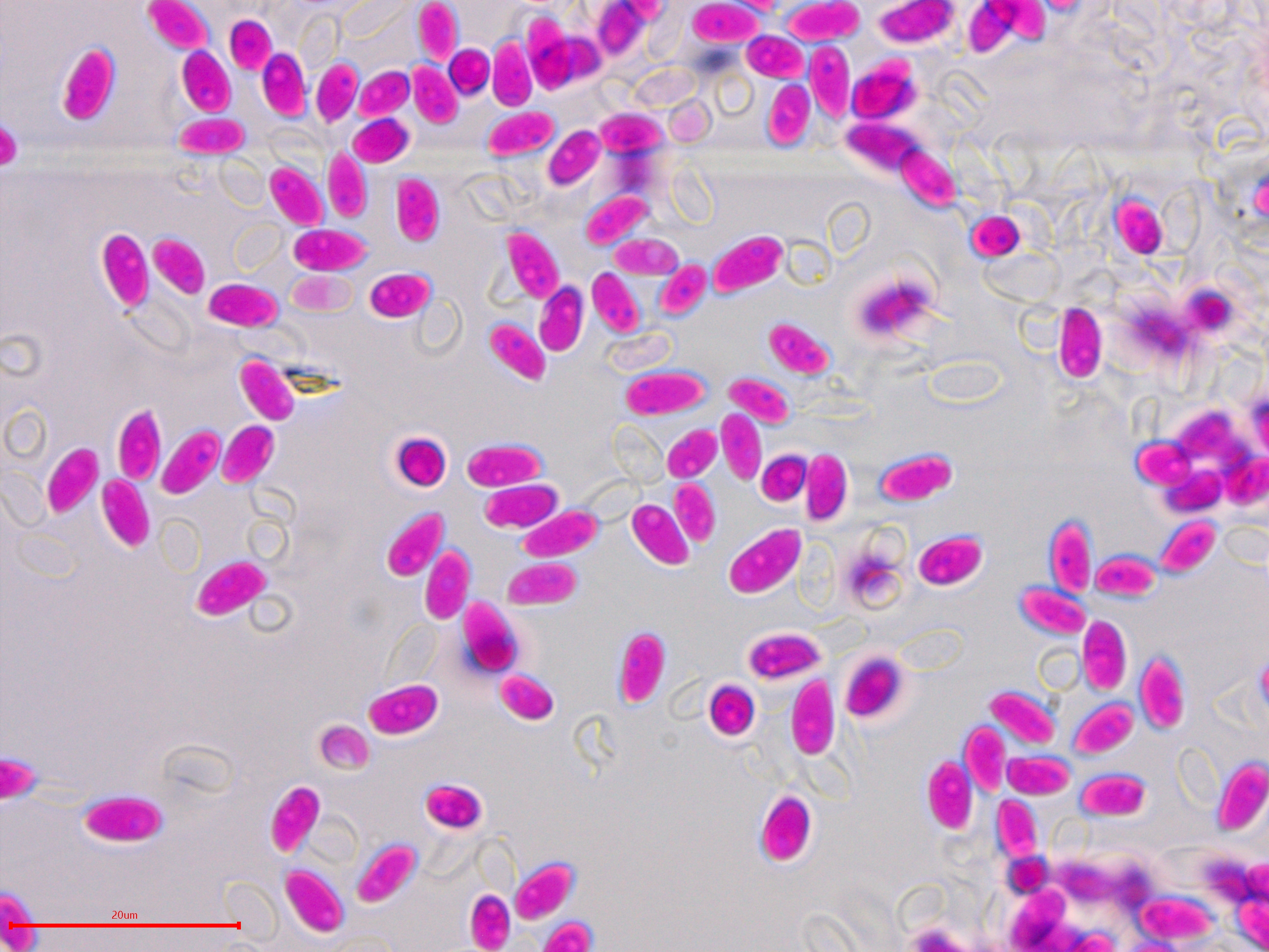
Cylindrical basidiospores.
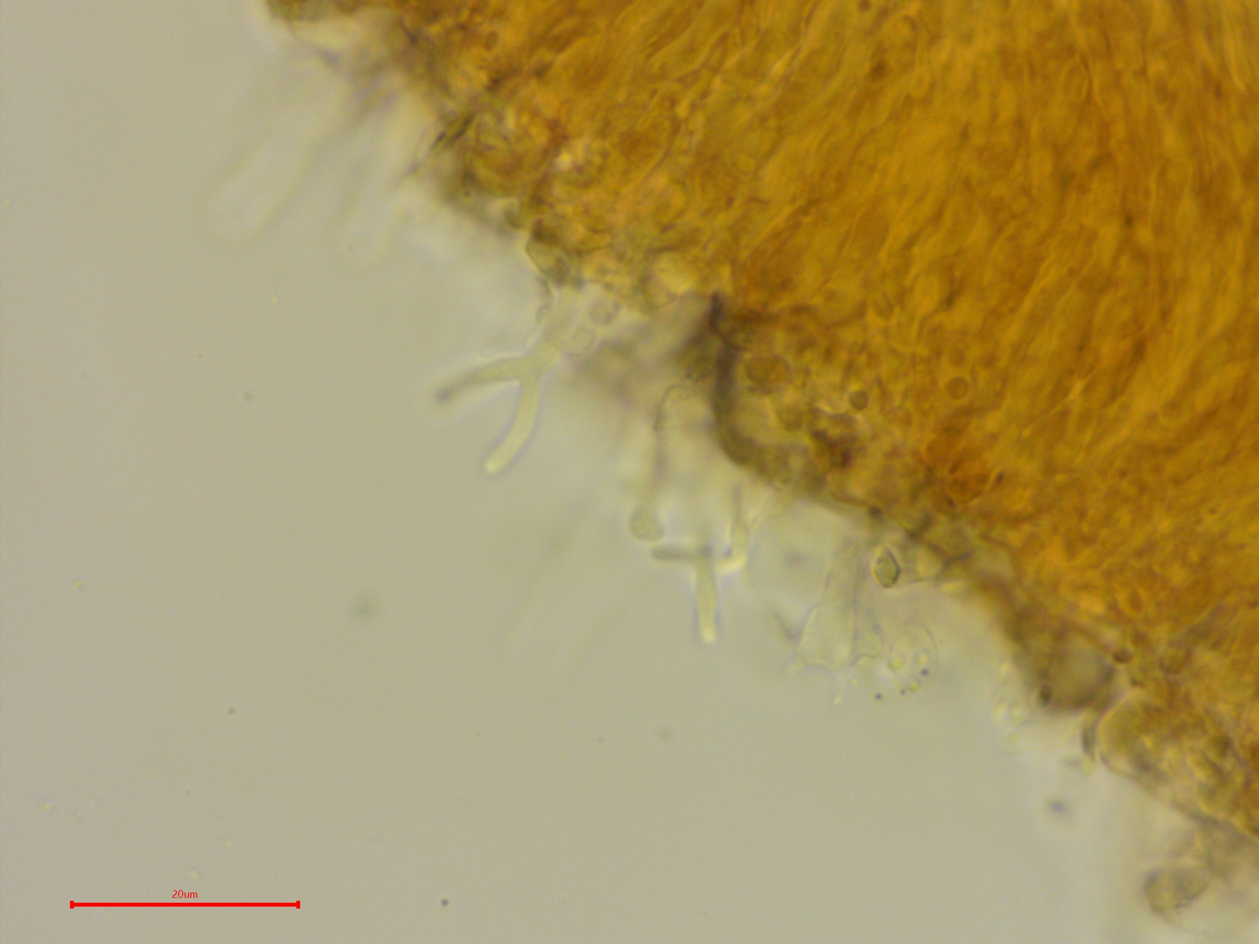
A look at projecting dendrohyphidia.
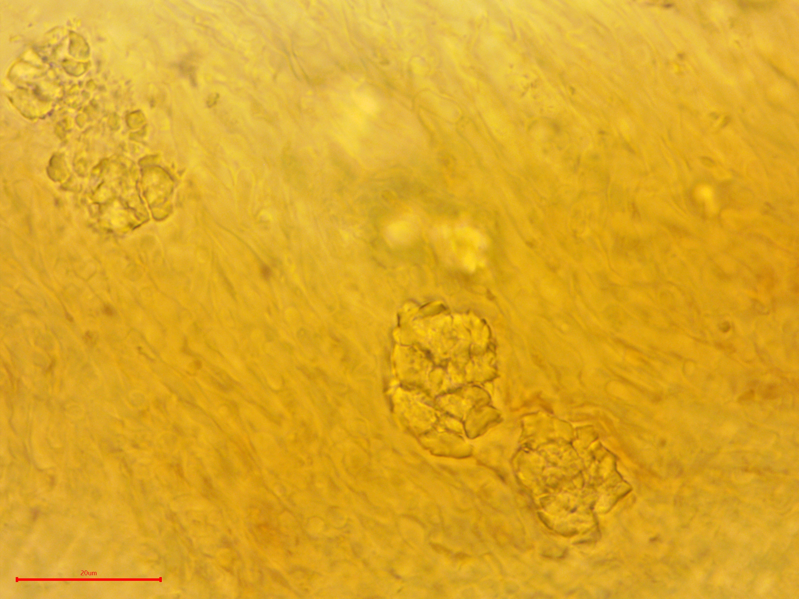
Granulated content was embedded in the hyphae.
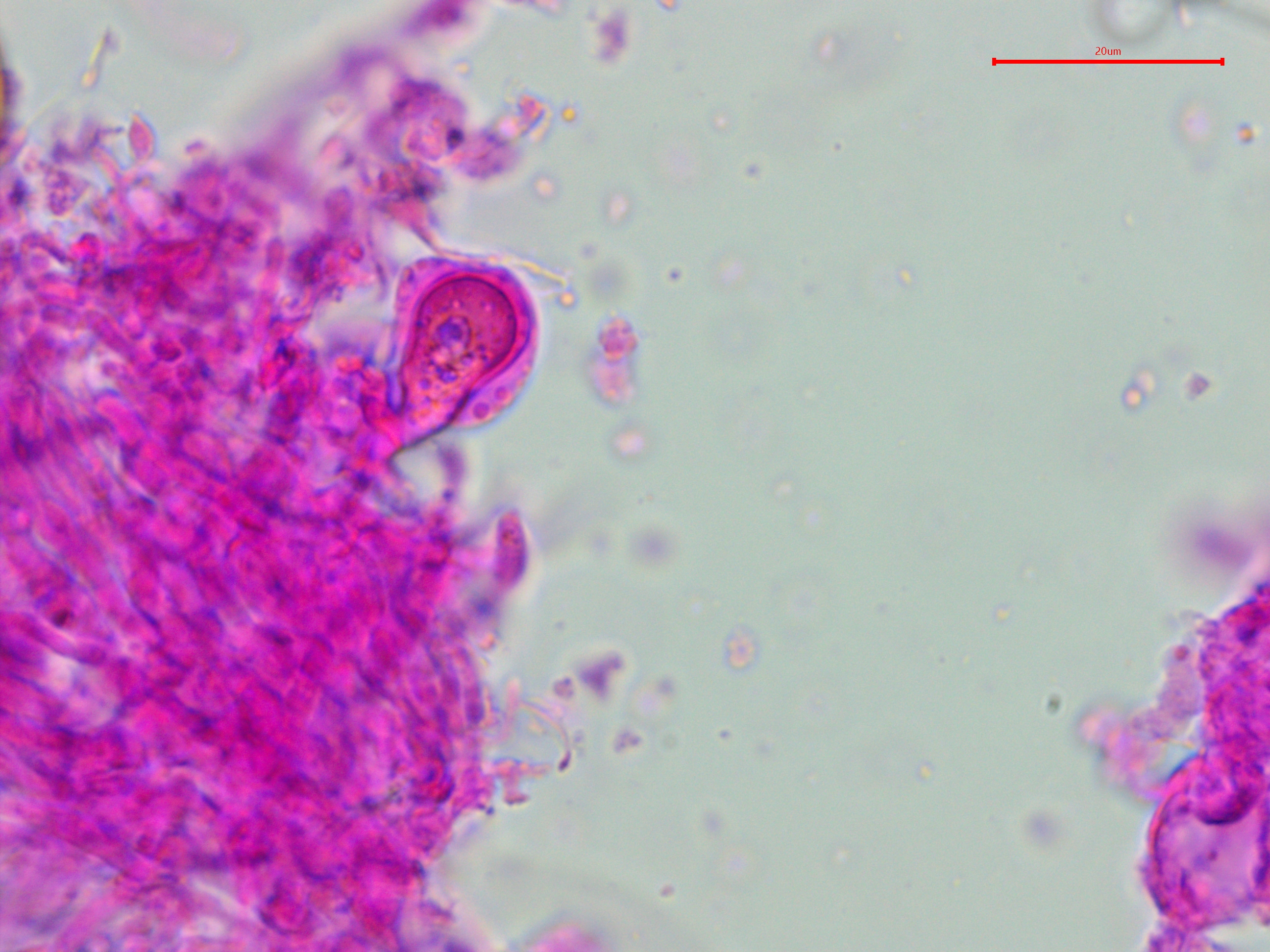
I only observed one of these strange structures.