Introduction to Crust Fungi
Overview
Crust fungi (or corticioid fungi) are of great ecological importance as well as morphological and evolutionary diversity. You can find them almost anywhere where there is wood, growing on the underside of a stick or log, on the bark of living trees, or even on the bottom of leaves, duff, and rocks. They make their living as saprotrophs, eating dead organic matter; or symbionts, gaining nutrition in intimate association with a living host. While mushroom hunters often scoff at crusts, fungal ecologists increasingly appreciate their importance for forest health and ecosystem functioning (Rosenthal et al. 2017, Anthony et al. 2022). Saprotrophic crusts are essential recyclers of nutrients in forest ecosystems and mycorrhizal crusts are often the dominant soil symbionts in coniferous forests. Some crusts even form mycorrhizal associations with orchids, meaning we wouldn't have these awe-inspiring flowers without humble crust fungi.
I'll be the first to admit that these aren't the flashiest of organisms. They are usually small and indiscrete, some nothing more than a sheen or different hue on the bottom of a decaying branch. And no, they are not edible. However, some crusts take on wild textures and colors whose evolutionary purpose defies explanation. If one dares to travel to the microscopic realms, they are greeted by truly bizarre structures like swollen oily cells, crystal-shrouded lances, and spiky clubs. To look into the microscope at a crust is to touch down on an extraterrestrial world with fractal geography and ornate constructions. In the study of crusts, you will broaden yourself as a mycologist and become acquainted with the taxonomic and microscpic nuances of almost all of Agaricomycotina.
Morphological Definition
While crusts often speak for themselves, mycologists do have a technical definition for what constitutes a corticioid fungus. According to Larsson (2007), corticioid fungi are basidiomycetes with effused, resupinate basidiomata; a smooth, merulioid, or hydnoid hymenophore; and holobasidia. However, I prefer the more expansive definition of Gorjón (2020), who defines crust fungi simply as "basidiomycete fungi in which basidiomes are generally resupinate". This means that they belong to the phylum Basidiomycota (the group of fungi that produce sexual spores on club-like structures called basidia) and generally grow flat along the bottom of a substrate. Beyond that, the hymenophore (the tissue that underlies the hymenium, or the spore-bearing surface) can be smooth or take on any number of configurations to increase surface area (bumps, folds, pores, or teeth); the fungus can be entirely flat or produce shelf-like caps, be thin and whispy, thick and rubbery, or whatever it likes. This broad definition naturally butts up against what mycologists refer to as stereoid fungi, a term used for fungi that produce caps with a primarily smooth hymenium, whether or not they also have some resupinate growth (resembling Stereum spp.); it also sometimes results in overlap with other form groups typically treated on their own such as polypores and hydnoid fungi. Finally, it includes fungi with septate basidia (heterobasidiomycetes), which were originally ignored by corticiologists because they were long considered a distinct taxonomic grup, but now we know they are integrated with the Agaricomycetes (e.g., Auriculariales, Cantharellales, Sebacinales) (Hibbett et al. 2014). The resupinate fungi in these orders certainly look like crusts — why exclude them only after discovering under the microscope that they have septate basidia? Ultimately, the best advice regarding what to consider a crust may be that of Henrici (2000): "It doesn't do to question too closely what is meant by a corticioid fungus... Any demarcation line will be arbitrary and the question of where to draw it is not one of importance."
Convergent Evolution of the Corticioid Form
Despite their apparent simplicity, a precise definition of crust fungi is difficult to formulate because they are a form group (defined by a vaguely similar morphology) rather than a monophyletic group (defined by a shared evolutionary history). In the past, all crust fungi were thought of as closely related species and were placed together in the family Corticiaceae — hence the term "corticioid", signifiying an affinity to fungi in the family Corticiaceae. Nowadays, we can sequence an organism's DNA to more accurately determine its relatedness to other species and this has drastically changed corticioid taxonomy. Crust fungi are actually distributed across most of the 21 orders in the the class Agaricomycetes, thoroughly intermixed with more popular mushrooms such as agarics, polypores, boletes, puffballs, and chanterelles (Binder et al. 2005, Hibbett et al. 2014, Zhao et al. 2017). Related to each of these very different types of mushrooms are crusts that, while looking superficially similar to one other, are actually only distantly related. This is shown below in Figure 1.
The contemporary diveristy of basidioimycete mushrooms is truly amazing, but if you were to go back far enough in time, crust fungi were all that there was. That's because Agaricomycetes descended from a crust-like ancestor (Sánchez-García et al. 2020). Over hundreds of millions of years, this group evolved extraordinary shapes, colors, and mechanisms of spore dispersal, but many lineages retained the crust form, and many others reverted back to this simple morphology (at least 96 times, according to the previously cited study). Crusts come and go again and again over evolutionary time like the kneading of dough. As a result, mycologists recognize a very large number of crusts (thousands of species) spread across more than 400 genera (Gorjón 2020) and 40 families (Larsson 2007). About 1000 species of crust fungi are known to occur in Canada and the United States alone (Ginns 1998). Given the fact that we have documented less than 10% of all fungi and that relatively little attention has been devoted to crusts, especially in the tropics, the number of species of crust fungi is most definitely far greater!
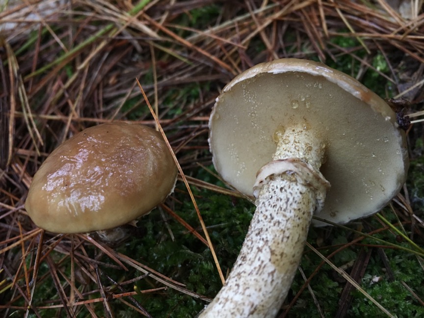
Boletales
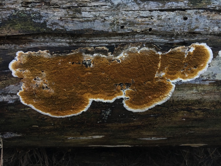
Polyporales
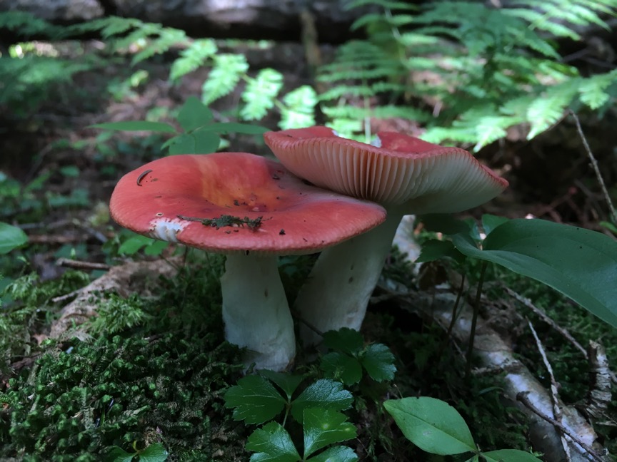
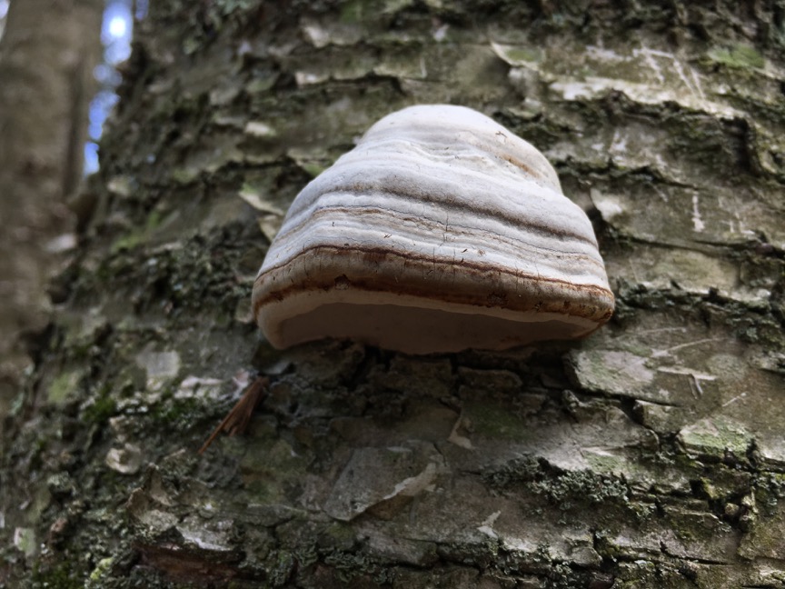
Russulales
Figure 1. Boletales, Polyporales, and Russulales are taxonomic orders that we typically associate with non-corticioid mushrooms (boletes, polypores, and agarics, respectively) but actually contain corticioid fungi. This shows that crust fungi are sometimes only distantly related, even if they have a similar morphology.
Mycologists Wanted
Why do I collect crusts and why should you? As a mycologist, I am curious about all the members of the Queendom Fungi. When you start foraging, you invariably run into crusts, and start to wonder, "who are you and what are you doing hiding under that log"? Not much accessible yet scientifically detailed information exists on the internet about crusts. I hope that CrustFungi.Com fills this niche so that interested individuals may better understand corticioid fungi. However, given that crusts are sadly understudied and underexplored, it will inevitably fall short. If you are intersted in discovering new species or having a large contribution to the documentation of the biogeography and ecology of fungi, whether as a community scientist or professional mycologist, crusts certainly have a lot to offer.
Collecting and Studying Crusts
Unlike the meditative wanderings of mushroom hunters, crusts are most easily discovered by meticulously searching a single area with lots of fallen branches. While crust fungi might be visible on the tops of the branches, you'll find the greatest diversity by picking the branches up and scanning their undersides. Log rolling is another fruitful method — you will be delighted by all the different organisms that grow and live on and under a large log, even when it is cold out. Success is maximized by looking under woody debris of multiple tree species, sizes, and stages of decay, as different crusts have different ecological and successional preferences, with newly fallen wood being the least interesting. The bark and dead branches of standing trees and shrubs are great places to find hardy species that don't mind drying out. More elusive species, like ectomycorrhizal crusts, often grow on the bottom of leaf debris and conifer duff, and are suprisingly prevalent on the underside of rocks (scree or lapidicolous fungi). Wooden debris in wet areas like bogs, marshes, riverbeds, and sea shores (yes, there are marine crusts!) sometimes harbor very unique species. Other specialty crusts can be found on diverse substrates such as lichens, herbaceous debris, and dead ferns, which are particulary small, delicate, and understudied. Finally, human constructions such as wooden fences, posts, railings, and bridges apparently also harbor small and rarely collected species (Hjortstam et al. 1973).
Macroscopic Traits
Many crusts are impossible to identify to species without microscopic study or DNA sequencing. I assume you have access to a compound microscope (one with up to 1000x magnification) in my comments below. If you do not have access to a microscope, some charismatic crusts can be identified by their macromorphology alone; check out the synoptic key or browse the photos on the species page to see if any are a good match. Regardless, succesful identification begins with close inspection of the crust and careful notetaking of its traits that are discernable by the naked eye. First, when and where did you find it? What is the substrate, the type of associated rot if a wood-decay fungus (only a few genera produce brown rot, which makes identification of brown-rot crusts easier), and nearby trees? If it is not a "typical" crust growing on the underside of a branch or log, this is important to note.
Then, what can you observe about the growth of the crust (open up the glossary in a new tab to read more about these and following terms)? Note the colors of the fruiting body, the hymenophore configuration, and its consistency. Make sure to also note what is going on along the expanding edge of the crust. Is the margin a distinct color from the rest of the fruiting body? How are the hyphae growing? Observation of these features is aided by a small hand lens or jeweler's loupe.
Once you have observed all these traits and photographed the crust in its fresh state, collect a sample for further study and preservation. A good knife is usually sufficient, but a small axe or saw may be necessary to handle hard wood. In whatever container you carry the specimen (plastic tackleboxes with adjustable walls work really well), try to place the crust in the same orientation that it was growing. Basidiomycete fungi will often reorient themselves so that their spores fall in the same direction as gravity. Try it out with gilled mushrooms — if placed horizontally, over a few hours they will completely bend over so that their gills are facing down. In the case of crust fungi, if placed upside down, the hyphae may start to grow into the spore-bearing surface resulting in a fuzzy appearance and challenging microscopy.
Spore Print
Back at home, you can try to get a spore print by putting the crust hymenium-side down on a piece of aluminum foil or a glass slide and placing a wet paper towel or cloth on top over night. The next morning, you may find a cloudy deposit of spores. Most crust fungi produce white spore prints, but you may collect some with spore prints that are more colorful. A spore print serves multiple purposes. For one, measuring spores will be a breeze: rather than tediously searching for spores in a squash mount, you have thousands that can be quickly scraped onto a slide and viewed and measured under the microscope. Second, the crust can be cultured from the spores for further study in the lab or to facilitate DNA sequencing. Third, some mycologists toss crusts that do not produce spore prints because these are likely to be infertile (without basidia or basidiospores), making morphological identification essentially impossible. Crusts should be dried at room temperature or in a dehydrator at low temperature (approximately 95 ˚F) as soon as possible after collection and acquiring a spore print. The specimens should be totally dried before storage, but should not be exposed to extreme temperatures in the dehydration process as this will modify or damage the microscopic structures.
Microscopic Traits
It is important to mention that you do not need to do microscopy on crusts when they are fresh (so-called vital taxonomy). In fact, crust species are described after being dried and therefore it is possible identification will be thrown off by unaccounted-for differences of the fungus in the fresh state. Depending on the thickness or hardness of the crust, a vertical section or scraping can be made. For more robust crusts, cut as thin of a section of the crust as possible with a single-edge razor blade, ideally under a dissection microscope. A good section will be so thin that it curls like a pencil shaving and include all layers of the fruiting body, from the substrate to the hymenium. Transfer the section to a drop of mounting medium on a glass slide, place a cover slip on top, and gently tap with a pencil eraser. For thin or otherwise insubstantial crusts, a scraping can be made by first dipping the razor blade in the mounting medium and then touching the razor blade to the crust to wet a small area. Scrape or cut a small amount of tissue and transfer to the drop on the slide. Various mounting media and stains can be used, and each is good for its own purposes (check out the glossary). If the section or scraping is thoroughly crushed by putting pressure on the cover slip, this is referred to as a squash mount.
Now that you have prepared a slide, observe the thickness and makeup of the different layers of the crust. This is best accomplished with a very thin, full section of the fruiting body that has not been crushed. Following that, make notes and measurements of the following traits, which are crucial for identification (and often easier to observe after thoroughly squashing the tissue): hyphal system and clamp connections; number of sterigmata on the basidia and basidia shape and size; basidiospore shape, size, ornamentation, and chemical reactions; and sterile structures such as cystidia. Often times multiple slides will have to be prepared from different parts of the crust to observe all these different features. It is also usually necessary to observe these traits in different media. For example, KOH stained with phloxine B is my default mounting medium, but Melzer's reagent is necessary to observe amyloidy of spores or other structures, and water may be necessary to accurately observe traits that dissolve in KOH (like halocystidia).
Sequencing Crusts
If not already, DNA barcoding is becoming a routine and standard part of the set of metadata attached to vouchered fungal specimens. Even for "amateur" scientists without access to a mycology lab, DNA sequencing is becoming more and more doable because of the ever decreasing cost of sequencing, the ability to assemble a barebones but functional lab at home, and access to third-party sequencing services, for example via Fungal Diveristy Survey. To be clear, "sequencing" refers to the "reading" of the order of base pairs that make up an organism's genetic code; "barcoding" refers to the sequencing of a specific genetic region that is useful for identification at the species level (like scanning a PLU code on produce in a grocery store).
What's so useful about sequence data, or why do we care to sequence the DNA of fungi? From an identification and phylogenetics perspective, each nucleotide position serves as a character that can be used in understanding how two specimens are related. So if you have a DNA sequence consisting of 700 nucleotides, you have 700 characters to work with, in addition to standard morphological ones. With morphology or chemistry alone, we have far fewer than 700 characters for comparison (try to come up with even 20)! While we might be able to compare and categorize morphological characteristics in our heads pretty easily, like color ("this specimen is blue and this specimen is red, they are probably not the same species..."), sequence data are more difficult and there's far more of them, so we rely on computer software to help us out. Over the long term, DNA data help tremendously with identification as species concepts and nomenclature evolve, and can be easily stored in databases for rapid data analysis. Finally, DNA data cut through morphological similarities to which we assign undue importance in species descriptions, ones that are either variable within a species or convergently evolved (do not reflect relatedness), to get at the true evolutionary history of a set of taxa.
While many DNA barcoding regions exist and all are useful in some circumstances, the universal fungal barcode is the internal transcribed spacer (ITS) region of the nuclear ribosomal DNA (rDNA) (Schoch et al. 2012). Ribosomal DNA is the DNA that is transcribed into ribosomal RNA, which is assembled into ribosomes, the molecular machinery that makes proteins in all living organisms. So every living thing has rDNA! The ITS region is a section of DNA that is transcribed along with the rDNA but is then cleaved out. In other words, it is a spacer region that never becomes a functional product. Mycologists have chosen this region as the universal barcode because it is relatively conserved at the species level but typically quite variable above that, given that it is not actually encoding for anything and can therefore gain many mutations without affecting the functioning of the cell.
A comprehensive tutorial on all the steps involved in sequencing fungi, from DNA extraction to bioinformatics, is beyond the scope of this introduction, but I offer a few crust-specific tips. For crusts, I like to place a small amount of tissue (a fragment of a white rice grain) each into a tube containing 5% Chelex and another containing 2X CTAB. With the Chelex tube, I first do a rapid but less reliable microwave DNA extraction method (Dörnte and Kües 2013). For the samples that did not extract succesfully using the microwave method, or for ones that I want high-quality DNA for whole genome sequencing, I use a chloform extraction protocol on the CTAB isolate (protocol available here). Professor Tom Bruns' method is to use an insect pin to pick up the smallest piece of tissue possible (just barely visible under a dissection scope) and to process that with Sigma-Aldrich's Extract-N-Amp kit. Since crusts are so two-dimensional, they are often heavily contaminated — a small amount of clean-looking tissue, along with the use of Basidiomycota-specific primers, typically result in the best amplification.
Resources
Literature
The first true compendium of crust fungi was Hyménomycètes de France by Bourdot and Galzin, published in 1928. Around 50 years later, The Corticiaceae of Europe in eight volumes (1973-1988) by Eriksson, Ryvarden, and colleagues became the new standard for corticiology. Today, Fungi Europaei, Volume 12: Corticiaceae s.l. (2010) by Bernicchia and Gorjón is the most useful reference available, even outside of Europe. While general treatments of North American crusts are relatively scarce, The Resupinate Non-poroid Aphyllophorales of the Temperate Northern Hemisphere (1980) by Jülich and Stalpers contains some good information and Stereoid Fungi of America (2010) by Ryvarden discusses many crusty fungi as well.
Online
For assistance in identification, general questions related to crust fungi, and community support, I recommend you join the group Crust Fungi and Polypores on Facebook. With over 3000 members across the globe, including some of the world's foremost corticiologists, it is an amazing forum to share and learn about crusts. Other useful online resources related to crust fungi include Elia Martini's very excellent website dedicated to Crusts and Jells, Patrick R. Leacock's MycoGuide of fungi of the Midwest and America (which does a superb job covering crusts), Michael Kuo's MushroomExpert.Com, the late Gary Lincoff's website with a gallery of 21 common crusts, Danny Miller's pictoral key to Pacific Northwest crusts, First Nature's British bracket and crust fungi gallery, a key to English crusts by the Hampshire Fungus Recording Group, and Sergio Gorjón's website containing keys, regional crust fungi checklists, and a directory of professional corticiologists.
References
Anthony, M. A., Crowther, T. W., van der Linde, S., Suz, L. M., Bidartondo, M. I., Cox, F., Schaub, M., Rautio, P., Ferretti, M., Vesterdal, L., De Vos, B., Dettwiler, M., Eickenscheidt, N., Schmitz, A., Meesenburg, H., Andreae, H., Jacob, F., Dietrich, H.-P., Waldner, P., … Averill, C. (2022). Forest tree growth is linked to mycorrhizal fungal composition and function across Europe. The ISME Journal, September, 1–10.
Binder, M., Hibbett, D. S., Larsson, K.-H., Larsson, E., Langer, E., & Langer, G. (2005). The phylogenetic distribution of resupinate forms across the major clades of mushroom-forming fungi (Homobasidiomycetes). Systematics and Biodiversity, 3(2), 113–157.
Dörnte, B., & Kües, U. (2013). Fast microwave-based DNA extraction from vegetative mycelium and fruiting body tissues of Agaricomycetes for PCR amplification. Current Trends in Biotechnology and Pharmacy, 7(4), 825–836.
Ginns, J. (1998). How many species are there? Folia Cryptogamica Estonica, 33, 29–33.
Gorjón, S. P. (2020). Genera of corticioid fungi: Keys, nomenclature and taxonomy. Studies in Fungi, 5(1), 125–309.
Henrici, A. (2000). An introduction to corticioid fungi. Field Mycology, 1(1), 12–19.
Hibbett, D. S., Bauer, R., Binder, M., Giachini, A. J., Hosaka, K., Justo, A., … Thorn, R. G. (2014). Agaricomycetes. In D. J. McLaughlin & J. W. Spatafora (Eds.), Systematics and Evolution: Part A: Second Edition (pp. 379–429).
Hjortstam, K., K.-H. Larsson, & L. Ryvarden (1988). The Corticiaceae of North Europe, Volume 1. Fungiflora, Oslo, Norway.
Larsson, K.-H. (2007). Re-thinking the classification of corticioid fungi. Mycological Research, 111(9), 1040–1063.
Rosenthal, L. M., Larsson, K.-H., Branco, S., Chung, J. A., Glassman, S. I., Liao, H.-L., … Bruns, T. D. (2017). Survey of corticioid fungi in North American pinaceous forests reveals hyperdiversity, underpopulated sequence databases, and species that are potentially ectomycorrhizal. Mycologia, 109(1), 115–127.
Sánchez-García, M., Ryberg, M., Faheema, K. K., Varga, T., Nagy, L. G., & Hibbett, D. S. (2020). Fruiting body form, not nutritional mode, is the major driver of diversification in mushroom-forming fungi. Proceedings of the National Academy of Sciences, 1–7.
Schoch, C. L., Seifert, K. A., Huhndorf, S., Robert, V., Spouge, J. L., Levesque, C. A., … Consortium, F. B. (2012). Nuclear ribosomal internal transcribed spacer (ITS) region as a universal DNA barcode marker for Fungi. Proceedings of the National Academy of Sciences, 109(16), 6241–6246.
Zhao, R. L., Li, G. J., Sánchez-Ramírez, S., Stata, M., Yang, Z. L., Wu, G., … Hyde, K. D. (2017). A six-gene phylogenetic overview of Basidiomycota and allied phyla with estimated divergence times of higher taxa and a phyloproteomics perspective. Fungal Diversity, 84(1), 43–74.